 Pet News


Summary
Dry eye, also known as keratoconjunctivitis sicca (KCS), is a common condition in dogs characterized by reduced tear production or poor tear quality, leading to inflammation of the cornea and surrounding tissues. Tears play a crucial role in maintaining the health of the cornea, and when their volume or quality is compromised, it can result in discomfort, corneal ulcers, and even blindness if left untreated. The tear film consists of watery, lipid, and mucin layers, each contributing to corneal moisture and protection.
Dry eye can have various causes, including immune-mediated attacks on tear glands, nerve disorders, developmental abnormalities, and underlying health conditions. Certain breeds, such as brachycephalics and dogs with chronic allergies, are more susceptible to dry eye. Clinical signs include redness, dull cornea, mucky discharge, excessive blinking, and pawing at the eyes. Diagnosis involves medical history, clinical signs, and tear measurement tests.
Treatment typically involves tear stimulants and tear replacements to stimulate tear production and provide corneal moisture, respectively. Regular monitoring and proper medication administration are essential, and in some cases, surgical intervention may be necessary. Early detection and intervention are crucial in managing dry eye and preserving a dog’s vision and comfort.
Bonnie, a 6-year-old, female, Cavalier King Charles Spaniel, presented to my clinic in August. Rita, who cares for Bonnie, reported that Bonnie had been noted to have red eyes on and off for approximately two months. More recently, however, she had developed a thick mucky discharge, initially from her right eye, but eventually from both eyes. Bonnie was squinting more often, which prompted Rita to have her examined by a veterinarian.
The antibiotic ointment prescribed by Bonnie’s veterinarian significantly reduced these signs. However, the redness, discharge, and squinting returned once the treatment concluded.
Upon further examination, her veterinarian performed a tear test and diagnosed Bonnie with dry eye. For the latest information about treatment options, Rita decided to consult with a veterinary ophthalmologist and happily this was me! Read on for more information about Bonnie’s treatment plan and response to treatment.
Overview - What is Dry Eye?
Dry eye, or “keratoconjunctivitis sicca (KCS)” as it’s formally recognized in the scientific community, is a common condition that occurs more frequently in dogs than many pet owners might realize. Dry eye is characterized by insufficient tear production and/or quality.
Tears are a vital aspect of maintaining the health of the cornea, the clear, outer layer of the eye. They create a moist environment necessary for the eye’s comfort and function while simultaneously acting as a protective barrier against foreign bodies and infectious agents.
Reduced tear production and/or quality can result in numerous complications and significantly diminish a dog’s quality of life. This overview examines the causes of dry eye, its signs, diagnostic methods, treatment options, long-term effects, and the prognosis for dogs dealing with this condition.
Wetness of the cornea is achieved by the tear film, or thin layer of tears that is spread evenly over the cornea by the eyelids. Dry eye occurs when the tear glands do not produce enough tears (quantitative dry eye), or the quality of tears produced are too poor to prevent tears from evaporating too quickly or spreading evenly across the cornea (qualitative dry eye).
Like a jellyfish, corneas must be always wet to maintain clarity and health. Tears have many important functions that apart from providing lubrication and comfort, maintain the overall health of the cornea by keeping the cells of the cornea and surrounding tissues healthy, washing away debris, and helping to fight bacteria.
When the tear volume or quality is reduced, the cornea becomes painful, appears dull, and if left untreated, can lead to serious complications such as corneal ulcers and even blindness.

The tear film is made up of three layers: a watery component (aqueous tear film) which is flanked by a thin fat (lipid) layer and a thin mucin layer [Fig 2]. The lipid and mucin layers help to keep the watery layer attached to the cornea, but also help to keep the watery layer from evaporating too quickly from the cornea.
The watery layer (aqueous layer) is made by two glands, the lacrimal gland, and the gland of the third eyelid [Fig 2]. The gland of the third eyelid is sometimes seen when it becomes swollen and pops up over the edge of the third eyelid. This condition is called prolapsed third eyelid gland (“cherry eye”) and will be discussed in another article.
Too little of the watery layer is the most common cause of dry eye. When the watery layer is reduced, the other two thinner layers (the lipid and mucin layers) continue to be produced normally or even increased, and this results in a thick gooey eye discharge [Fig 3]. In contrast, the lipid and mucin portions of the tear film are made by small glands that are found along the edges of the eyelids and by the conjunctival tissue (the pink slippery tissue that surrounds the entire eye). [Fig 2].
A reduction in one or both layers results in a poor quality of tear whereby the watery layer is either not spread evenly across the cornea or allows the watery layer to evaporate too quickly from the surface of the cornea.
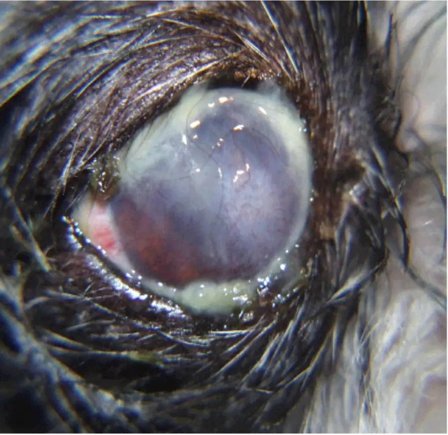
Photo Credit Marnie Ford, DVM.
What are the Causes of Dry Eye?
There are many causes of dry eye in dogs. The most common cause of dry eye in dogs occurs when the body’s immune system attacks the gland(s). This form of dry eye is called immune-mediated dry eye. In such a situation, the dog’s immune system incorrectly identifies its own tear glands as threats, leading to their destruction and subsequent reduction in tear production. It is a complex and unfortunate process, but one that’s critical to understanding the condition.
Other causes of dry eye include disorders of the nerve which normally stimulates the tear gland to produce tears (neurogenic dry eye), developmental abnormalities whereby the gland(s) did not develop (this is most common in Yorkshire Terriers), prior medical procedures (eg removal of the third eyelid gland, local radiation therapy, general anesthesia (usually this is transient)), genetic predisposition (eg Cavalier King Charles Spaniel, West Highland White Terrier), underlying health conditions such as hypothyroidism, diabetes, and Cushing’s Syndrome, trauma to the gland(s) or nerves, as a consequence of some medications (eg oral sulfonamide drugs, atropine eye drops) or prior infections (eg canine distemper virus infection, leishmaniasis, chronic eyelid inflammation).
Which dogs are most commonly affected by Dry Eye?
Any breed of dog can develop dry eye but flat-faced dogs (brachycephalics) such as Pekingese, Pugs, Boston Terriers, Bulldogs, Lhasa Apso, and Shih Tzu’s, dogs with chronic allergies (eg West Highland White Terriers), or other breeds such as the English Springer Spaniel, American Cocker Spaniel, Bloodhouse, Cavalier King Charles Spaniel, Miniature Schnauzer, Samoyed, and Yorkshire Terrier are most often affected.
Dry eye most commonly affects middle aged to older dogs. However, when caused by malformed or absent gland tissue, dry eye will often develop before the age of 2 years.
Advertisement
What are the Signs of Dry Eye in Dogs?
Dogs suffering from dry eye may display a wide range of signs that vary depending on severity and duration of the condition. The signs of dry eye can include redness of the normally pale/light pink tissue around the eye (the conjunctiva), a dull looking cornea, thick sticky discharge, excessive blinking or persistent squinting, and pawing at the eye(s).
In some cases, dogs may develop corneal scarring and/or pigmentation, and may develop corneal ulceration, which can cause severe pain and may require surgery to treat. With persistent mucky discharge, the eyelids are often noted to develop a crusty buildup of material.
Typically, these signs develop in both eyes, but one eye may appear worse than the other. When caused by lack of gland formation, trauma, radiation, or gland removal only one eye is usually affected.
It’s important to note that these signs can also be present in other eye conditions, which makes it all the more necessary to consult with a veterinarian for a correct diagnosis. In the early stages of the disease, these symptoms might only occur intermittently.
However, as the disease progresses, symptoms are likely to become more constant. Unfortunately, by the time symptoms become persistent, irreversible damage to the tear glands and or cornea may have already occurred, resulting in poor medical response to treatment and reduced vision or even blindness. This underlines the importance of regular veterinary check-ups and early intervention.
How is Dry Eye Diagnosed?

Photo Credit Marnie Ford, DVM.
Diagnosis of dry eye is based on medical history, clinical signs, and measurement of tear volume and evaluation of tear quality. Diagnosis of quantitative dry eye (when the watery layer is low) is by the Schirmer Tear Test (STT). This test is simple and painless and can be performed by most veterinarians [Fig 4].
To test for this, a sterile calibrated strip of paper (a Schirmer tear strip) is placed under the lower eyelid to contact the surface of the eye for 1 minute. The tear strip has 1 mm calibrations which indicate the level tears that are absorbed over the 1-minute testing time. Normal tear production is greater than 15mm of tear wicking on the strip per minute.
Tear production less than 10 mm/min is not normal and supports a diagnosis of quantitative dry eye [Fig 5]. If this test indicates a normal level of tear production but the clinical signs of dry eye are present (dry looking cornea, mucky discharge, redness), a visit to a veterinary ophthalmologist may be required. A veterinary ophthalmologist can diagnose the qualitative form by using special stains on the cornea.
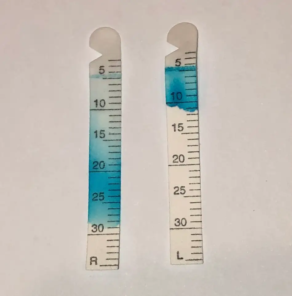
Photo Credit Marnie Ford, DVM.
Because dry eye can expose the sensitive cornea to additional problems, additional tests that are often performed include corneal staining to check for corneal ulcers, intraocular pressure (IOP) to determine if inflammation or glaucoma is present, and examination of the tear ducts to ensure that the tears are able to drain properly.
The STT is often performed every 3-6 weeks after starting treatment to ensure that the medication(s) and treatment plan is working well for your dog. Improvement in comfort and reduction of mucky discharge is usually noted within the first 2-3 weeks after starting treatment and once an adequate tear production has been achieved, less frequent monitoring of tears is usually recommended.
In most cases, treatment will be required for the life of your dog; however, the frequency and number of medications used is often greatly reduced as the tear production and/or quality increases. In some case, treatment can be stopped if the underlying cause of dry eye is stopped or controlled (e.g. use of some medications are stopped, the effects of prior anesthesia have worn off, treatment for hypothyroidism is started).
Advertisement
How is Dry Eye Treated?
Treatment for dry eye in dogs typically involves the use of two different kinds of medication. The first stimulates tear production (lacrimostimulant) and the other provides moisture (lacrimomimetic). The goals of all treatment for dry eye are to provide corneal comfort, clarity, and protection.
Remember, corneas always require moisture to remain clear and healthy. Without timely and appropriate treatment, dry eye can result in severe complications. These can include keratitis (which is a painful inflammation of the cornea), corneal ulcers, or scarring, which can interfere with vision.
Over time, untreated dry eye can even lead to a complete loss of vision. It’s therefore crucial that dry eye is detected and treated as early as possible to minimize the risk of these complications. Again, corneas can be thought of like a jellyfish…when found out of water they quickly develop a grey, unhealthy appearance that is easily damaged.
When diagnosed early, your dog has a very good chance of maintaining vision and comfort. Tear stimulants are prescribed for immune-mediated dry eye or neurogenic dry eye.
Immune-mediated attack on the lacrimal gland(s) is the most common cause of dry eye in dogs and as such, medications that work to stop or reduce the local immune-system from attacking the tear gland(s) are most prescribed. This consists of a medicated drop or ointment applied directly to the affected eye(s) usually once or twice a day.
Most commonly, cyclosporine (such as brand name Optimmune®ointment) or tacrolimus is prescribed as a drop or an ointment to stimulate tear production. These immunomodulating medications are often compounded for topical use, and when applied to the eye, keep the immune system from attacking the glands that produce the watery portion of the tear film. By inhibiting immune-mediated attack on these glands, the glands can continue to produce the watery layer of the tear film. These medications often take several weeks to work to their full capacity and consistency in application is required.
Alternatively, neurogenic dry eye is caused by reduced or absent nervous input to the gland(s). Neurogenic dry eye is most commonly caused by physical damage to the nerve or gland (e.g., injury from a bite to the head or damage to the head).
Treatment for neurogenic dry eye consists of administration of a drug that mimics the nerve that normally innervates the gland to stimulate tear production. Pilocarpine (brand name Isopto Carpine®) is a parasympathomimetic (i.e., mimics the parasympathetic nervous system) that unlike cyclosporine or tacrolimus, is a commercial eye drop that is administered by mouth.
Lacrimomimetics (tear replacements), or treatments that provide moisture to the cornea are critically important in the early weeks following diagnosis of dry eye since the tear stimulating drops often take several weeks for tear function to increase. These medications need to be administered frequently, often several times a day, due to the rapid rate at which tears naturally evaporate or are cleared from the surface of the eye.
Artificial tears provide vital functions such as moisturizing the surface of the eye, improving comfort, and helping to flush mucky accumulation away from the eye.
Unfortunately, these watery drops do not last long on the eye and as such, thicker lubricants are also required. Thicker and longer lasting lubricants are available in the form of oils, gels, or ointments.
Your veterinarian or pharmacist will be able to assist you with finding many options available without a prescription. Most dogs are treated initially with a tear replacement every two to six hours, depending on severity of the dry eye. The need for tear replacement will become less and less as the natural tears are increased.
In addition to treatment with tear stimulants and tear replacement medications, some dogs will also require additional medications. These may include antibiotic drops and or anti-inflammatory medications. These medications are used to treat underlying infection and or inflammation that may have been caused by the presence of the mucky discharge and the reduced natural cleaning ability of tears. These additional treatments are often temporary.
When tear glands cannot be stimulated to produce tears, when patients are born without glands at all, or if you are unable to administer your dog’s eye medication, surgery may be necessary. Surgery can be done to reroute one of the many salivary ducts entering the mouth, to the inside of the lower lid to create a path for saliva to reach the eye! This procedure is called a parotid duct transposition (PDT) and results in a wet eye, albeit with saliva and not tears. Unlike tears, saliva pH is basic and contains minerals.
Surgery is not an option for every dog, but the veterinary ophthalmologist who can perform the surgery will determine if your dog is a good candidate for this procedure. This procedure has significant risk of complication so it should not be pursued unless all other treatments have failed.
Complications that can occur include blockage of the salivary duct, overproduction of saliva, underproduction of saliva, or mineral deposition onto the cornea. This surgical procedure, while providing moisture to the cornea, is not curative and can lead to chronic problems that require continued use of topical and/or oral medications.
Regardless of the medication(s) recommended, maintaining clean short hair around the eyes will not only help your dog feel better, but will help to reduce the potential for corneal damage that can occur from rubbing at the eyes with a paw.
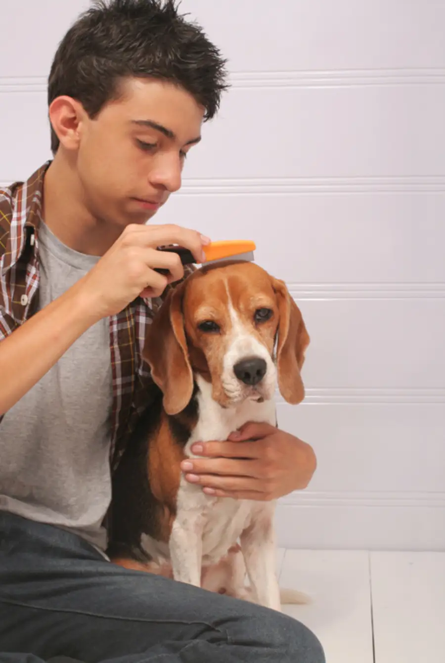
A warm wet facecloth applied to the closed eyelids (like a compress) for several minutes will help to soften any crusty debris that can then be combed out of the hair using a flea comb or other fine-toothed comb [Video 1]. Your veterinarian can demonstrate the correct way to administer medications and clean the hair around your dog’s eyes.
What is the Prognosis for Dogs with Dry Eye?
While dry eye is a chronic condition that requires ongoing management (usually lifetime), many dogs with this condition can lead relatively normal and comfortable lives with the right care and treatment regimen. Regular veterinary check-ups are necessary to monitor the progression of the disease and adjust the treatment plan as necessary.
The key to managing dry eye is consistency. Regular administration of medications, along with any necessary lifestyle adjustments, can help to control symptoms and minimize the risk of complications.
It’s important to remember that every dog is different, and a treatment regimen that works well for one dog may not necessarily be as effective for another. Therefore, it’s crucial to work closely with your veterinarian or veterinary ophthalmologist to develop a customized treatment plan for your dog.
So, what happened to Bonnie? During Bonnie’s initial visit with me, I confirmed her diagnosis of quantitative dry eye, the most common form of the condition. Fortunately, there are several effective treatment options for this type of dry eye. As Bonnie’s tear levels were mildly low, at exactly 11 mm/min, I prescribed Optimmune® and a corneal lubricant.
Six weeks later, Bonnie’s tear levels had returned to the normal range at 21 mm/min. I discontinued the lubricant as her tear production was normal and there was no longer any mucky discharge, redness, or discomfort.
Her next eye examination will be in six months. I am optimistic that with consistent treatment with tear-stimulating medication the rest of her life, her tear levels will remain within the normal range. If Bonnie’s tear levels or tear quality decrease in the future, other tear stimulants such as cyclosporine at a higher concentration or tacrolimus can be prescribed for her.
Advertisement
Conclusion
Dry eye is a common condition in dogs that can be managed effectively with the right knowledge, care, and treatment. When left untreated, dry eye results in discomfort and potential vision loss.
While prevention of dry eye is not always possible, early detection through regular veterinary check-ups, and monitoring for increased mucky discharge and redness by you at home, the crucial steps needed for early management and prevention of long-term problems are taken. With early intervention and diligent treatment, your dog can live a happier life with comfortable, clear eyes!
By Marnie Ford PhD, DVM, Diplomate ACVO
Dr. Ford graduated from the Ontario Veterinary College in 2000 after completing a Bachelors in Zoology at the University of British Columbia and a PhD in Physiology at Monash University in Australia. Following a rotating small animal internship at the University of Minnesota in 2001, Dr. Ford achieved Diplomate status from the American College of Veterinary Ophthalmologists in 2006 after completing a 3-year residency in Comparative Ophthalmology at the University of Missouri. After co-founding a private referral hospital, West Coast Veterinary Eye Specialists, in New Westminster (2004-2013) she opened Pacific Animal Eye Specialty services, providing mobile medical and surgical ophthalmologic care to patients in the Lower Mainland, Vancouver Island, and the Interior of BC (2013-2019). In 2019, she returned to Australia where she worked full-time in both medical and surgical ophthalmology as well as resident training at Animal Eye Care in Melbourne Australia. In 2023, Dr. Ford returned to Vancouver to provide locum services to ophthalmologists across Canada.