 Pet News


Summary
As veterinary medicine advances, our patients are living longer and as such, have more age-related problems. It is important to distinguish normal age-related changes from disease. Ophthalmic examination of the senior patient requires a slow approach that because of sore joints and weakened muscles allows them to have frequent position changes and slip-free surfaces.
Senior cats may benefit from the security of being gently wrapped in a towel for easier manipulation. Key age-related ophthalmic changes include modifications to the eyelids, cornea, iris, and lens, notably lenticular sclerosis, which involves the normal hardening of lens layers that impairs close vision and light sensitivity but does not obstruct vision like a true cataract. Accommodative measures such as increased lighting can significantly improve the quality of life for these animals.
Sam, a yellow-coated tabby cat, may be entering his golden years, but his spirit remains as bright as ever. Despite age-related ophthalmic changes, including lenticular sclerosis, Sam continues to explore his world with determination.
His owner, Mrs. Weatheringay (better known as Mrs. W), noticed Sam’s hesitance in dimly lit environments. Concerned, she consulted their veterinarian, who confirmed Sam’s condition. Determined to enhance Sam’s quality of life, Mrs. W embraced the vet’s recommendations.
Armed with knowledge about Sam’s visual challenges, Mrs. W transformed their home. Night lights illuminated Sam’s favorite spots, ensuring he navigated safely. Outdoors, Sam sported stylish visor, shielding his sensitive eyes from the sun’s glare. Sam tolerated the visor well. He knew that it made him feel better when outdoors.

Introduction
Ophthalmic changes noted in senior dogs and cats most commonly affect the eyelids, cornea, iris, and lens. Lenses are made up of layers of clear lens fibers arranged somewhat like an onion. Over time, new lens layers compress (sclerose) pre-existing layers. These hardened lenses are called sclerotic lenses and referred to as lenticular sclerosis.
Lenticular sclerosis is sometimes referred to as “senile cataract,” because both lens changes give a grey appearance to the lens. However, this is a misleading term as a true cataract is an opacity that obstructs vision through all, or part of the normally clear lens. In contrast, a sclerotic lens does not typically obstruct vision but does reduce the lens’s ability to focus.
As lenses naturally age, they become harder and greyer and as such, less plastic. Consequently, the lens is less able to help the eye to focus on near objects. This is one of the reasons that you and I require reading glasses as we age!
Advertisement
Lenticular Sclerosis
Lenticular sclerosis not only reduces near vision and focusing, but also increases the need for more light. For these reasons, many older animals find dusk the most challenging time to navigate their environments; often tripping on objects or becoming more hesitant on stairs. The visual deficits are more obvious in dogs than in cats because cats have some advantages that help them to see better at night. (Please see my article on “How and What Cats See” for further information.) To help your pet navigate in dim conditions, use of night lights, motion lights, or carrying a flashlight when out for walks in the evening can make a big difference!
Confirmation of lenticular sclerosis (density) versus cataract (opacity) is made if the retina at the back of the eye can be viewed through all parts of the lens. To examine the back of the eye fully, your veterinarian or veterinary ophthalmologist will dilate the pupils using a short-acting eye drop called a mydriatic.
By shining a bright light through the dilated pupil, the sclerotic lens will often be seen as two concentric circles, the central, older nucleus being denser than the outer, younger and less compact cortex. Additionally, the retina in the back of the eye can be seen using a hand-held lens and light source. Older patients with sclerosis may also develop cataracts as well. A thorough eye examination is essential when cataracts begin to appear to determine if the patient will be a candidate for cataract removal surgery. Lenticular sclerosis does not progress to blindness.
Advertisement
Iris Atrophy

Iris atrophy is the progressive thinning of iris tissue to result in a scalloped appearance of the pupillary margin or a moth-eaten appearance of all/parts of the remaining iris tissue. Iris atrophy also commonly results in weakening of the iris muscles and results in iris tissue that cannot close (constrict) fully to remain enlarged (dilated). Iris atrophy may not develop at the same rate between the two eyes, and anisocoria is commonly noted.
Anisocoria from naturally occurring iris atrophy needs to be differentiated from diseases such as glaucoma, Horner’s syndrome, uveitis, nerve abnormalities, or application or ingestion of drugs/medications. While not vision threatening, iris atrophy causes increased sensitivity to light and squinting secondary to overwhelming brightness on sunny days. When irritation is noted, providing your pet with shady areas to play or go for walks is recommended. Tinted goggles for both dogs and cats are available if required for outdoor activity.
Eyelid Tumors

Eyelid tumors such as meibomian gland adenomas or other non-specific growths are most frequently noted in the older dog patient but occasionally these tumors can occur in cats. These tumors are usually benign and have a tendency to bleed. They can spontaneously fall off the skin and later regrow, or they can cause local eyelid swelling.
Removal of these masses by a simple surgery called a wedge resection is recommended before their growth exceeds ¼ the eyelid length, corneal irritation is noted, or if they bleed repeatedly. Alternatively, the masses can be treated by freezing the tissue to selectively kill tumor cells (cryosurgery). If these masses become larger than ¼ the eyelid length, removal can sometimes be more complicated, so early examination is recommended.
Advertisement
The Cornea

The cornea, when healthy, is clear thanks to perfect organization of very thin corneal collagen layers, and its water-barrier defenses. Watery tears on the outside of the eye and aqueous humor on the inside of the eye are denied entry into the normally dehydrated cornea by its outermost layer, the epithelium, and innermost layer, the endothelium. Without the presence or proper function of these layers, fluid can saturate the cornea to thicken it and turn it grey.
The epithelium is made up of 8-12 cell layers. The cells in the outermost layer of the epithelium are constantly being shed and renewed – much like skin. Focal loss of these cell layers is not uncommon when the eye is injured. When all layers of the epithelium have been lost an ulcer is created and fluid can enter the cornea at this site to cause focal edema (fluid accumulation) and grey discolouration. When the ulcer is healed, the newly formed cells of the epithelium (epithelial cells) can resume their function of helping to maintain corneal dehydration and clarity.

The endothelium is a single cell layer thick and made up of endothelial cells which do not renew. These cells, which are in direct contact with the aqueous humor, contain pumps that actively pull fluid out of the cornea to maintain dehydration. Loss of endothelial cells because of normal aging results in progressive corneal greying (corneal edema) with a distinct “honeycomb” appearance.
These lost endothelial cells are not replaced. Sometimes, a variation of endothelial degeneration can also occur in younger dogs – most commonly the Boston Terrier and Chihuahua. When the loss of endothelial cells has been severe, such as in advanced age, or accelerated cell loss because of trauma or disease (uveitis, glaucoma), too much fluid accumulates within the cornea to form small blisters or bullae.
Bullae formation can predispose a cornea to ulcer formation which in turn will worsen the edema by increasing fluid access through the epithelial route. In this state, corneal barriers and dehydration can be difficult to reestablish. While endothelial cell loss cannot be halted medically, the amount of fluid accumulating within the cornea may be slowed with the consistent application of hypertonic saline solution or ointment. The increased salt content of these medications helps to draw water out from the cornea.
With consistent use, partial clearing of the cornea may be noted, and the frequency of corneal ulcer formation reduced. Like looking through frosted glass, the presence of dense corneal edema will impact vision. Because treatment options are so different, corneal edema associated with endothelial degeneration must be differentiated from corneal edema caused by glaucoma or uveitis.
As previously stated, the cornea is normally clear because the cornea is dehydrated and the collagen layers are tightly stacked with great precision. With age, the clear corneal laminations of collagen become microscopically separated—like a stack of old paper vs those of a new textbook. Blood vessels present in the cornea from a past injury or inflammation can leak a minute amount. When leakage occurs between two microscopically separated “sheets” of collagen, the pattern of blood will be that of a fan, as seen in figure 1. This is called stromal hemorrhage. While sometimes dramatic in appearance, this benign finding is not painful and does not typically require treatment.
Eyelid Conformation
Eyelid conformation is not immune from the perils of aging. Rolling in of the lower eyelid (entropion) is usually thought to occur in young animals with overly long eyelids. However, entropion is also frequently identified in older animals, especially cats, with weight loss.
Advertisement
Enophthalmia
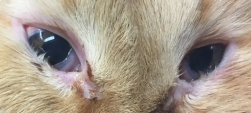
Loss of fat from behind the eyes results in eyes that sit deeper within the orbit. This is called enophthalmia and results in reduced eyelid support. Consequently, the eyelids roll inward, and the hair of the eyelid contacts and irritates the sensitive cornea. This irritation results in further withdrawal of the eye into the orbit and perpetuation of the cycle. Surgical correction of eyelid entropion is recommended to improve conformation and comfort. Surgery may involve cutting the eyelid tissue as described in the article Entropion in Dogs or injection of dermal filler to plump out the eyelid to reduce/eliminate the entropion. If surgical correction of the eyelids is not possible, application of a thick corneal lubricant several times daily is recommended for comfort and protection of the corneas.
Indolent Ulcers (Chronic Corneal Erosions)
Indolent ulcers (also known as chronic corneal erosions) are a type of slow-healing superficial ulcer that can be present in older dogs and cats. These ulcers are slow to heal because of an inability for the epithelium to stick to the lower layer of the cornea (the stroma). For this reason, they are considered mechanical. To heal these stubborn ulcers, several procedures can be performed by your veterinarian or veterinary ophthalmologist to encourage the epithelial tissue to stick to the underlying stromal tissue.
These procedures are: corneal debridement using dry cotton-tipped applicators or a diamond burr (diamond burr debridement); grid keratotomy or striate keratotomy using a needle to scratch the cornea; or as a last resort, a superficial keratectomy which involves removing the superficial ¼ to 1/3 of the cornea. Each of these procedures, while seemingly counterintuitive to healing, promote a rich infiltration of blood vessels into the cornea to promote healing but the corneas can also heal well without a vascular response. A topical broad-spectrum antibiotic and hypertonic saline therapy are recommended. (Please see my article in Pet Health Harbour entitled “Corneal Ulcers – An Overview” for further information.)
Advertisement
Conclusion
Effective management of age-related ophthalmic changes in pets during their senior years is crucial for enhancing their comfort and functionality. Understanding the distinct characteristics of conditions like lenticular sclerosis and implementing supportive environmental modifications can significantly aid in navigating the challenges associated with pet aging.
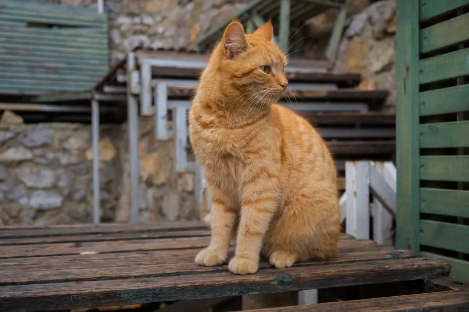
With these adjustments, Sam’s zest for life remained undiminished. His once tentative steps regained their confidence, as he ventured through familiar spaces with ease. Whether basking in the sun or chasing shadows, Sam’s joy knew no bounds.
Through understanding and adaptation, Sam and Mrs. Thompson showcased the resilience and bond between human and feline. Together, they embraced Sam’s senior years, proving that age may alter sight, but it can never dim the spirit of adventure.
By Marnie Ford PhD, DVM, Diplomate ACVO
Dr. Ford graduated from the Ontario Veterinary College in 2000 after completing a Bachelors in Zoology at the University of British Columbia and a PhD in Physiology at Monash University in Australia. Following a rotating small animal internship at the University of Minnesota in 2001, Dr. Ford achieved Diplomate status from the American College of Veterinary Ophthalmologists in 2006 after completing a 3-year residency in Comparative Ophthalmology at the University of Missouri. After co-founding a private referral hospital, West Coast Veterinary Eye Specialists, in New Westminster (2004-2013) she opened Pacific Animal Eye Specialty services, providing mobile medical and surgical ophthalmologic care to patients in the Lower Mainland, Vancouver Island, and the Interior of BC (2013-2019). In 2019, she returned to Australia where she worked full-time in both medical and surgical ophthalmology as well as resident training at Animal Eye Care in Melbourne Australia. In 2023, Dr. Ford returned to Vancouver to provide locum services to ophthalmologists across Canada.