

Advertisement

Eyes

Diseases & Conditions   |   Published on June 11, 2024

Ophthalmic Examination in the Adult Patient

Marnie Ford

Marnie Ford PhD, DVM, Diplomate ACVO

Summary

This article “Ophthalmic Examination in the Adult Patient” discusses common and uncommon ophthalmic conditions in adult dogs, primarily focusing on conditions that appear in the second and third quarters of a dog’s life. It covers a range of issues including cataracts, corneal degeneration, corneal dystrophy, glaucoma, and blindness due to retinal changes.

The inherited and acquired origins of these conditions are emphasized and their symptoms, progression, and the necessary evaluations and treatments discussed. Early detection and management of these conditions are critical particularly with potentially blinding conditions like cataracts and glaucoma.

In the quaint, leafy neighborhood where Silvia lived, everyone knew Wiley, a mature, somewhat overweight, and lately lethargic Golden Retriever with a contagious zest for life. However, his usual exuberance had diminished.

Wiley’s eyes began showing progressive redness and tearing, and Silvia noticed a worrying change in the color of his corneas. Concerned, she spent evenings researching and realized these could be signs of corneal degeneration, a condition she had never encountered before.

Taking no chances, Silvia brought Wiley to their trusted vet, Dr. Jensen. After a thorough examination, which included some blood tests, Dr. Jensen determined that Wiley had corneal degeneration linked to fatty deposits—exacerbated by his weight—rather than corneal dystrophy. The diagnosis was further complicated by an underlying hypothyroidism, contributing to Wiley’s weight issues.

Dr. Jensen prescribed tacrolimus eye drops to help control the inflammation and assist in removing some of the deposits, a corneal lubricant to keep Wiley’s eyes moist, and oral thyroid medication to manage his thyroid levels. Silvia absorbed every word, her heart heavy but filled with hope, as she learned how to administer Wiley’s new treatments.

Introduction

The adult patient, defined as the second and third quarter of life, can present with a wide variety of inherited or acquired ophthalmic changes that commonly include cataract formation, corneal dystrophy, corneal degeneration, glaucoma, keratoconjunctivitis sicca (KCS), and Golden Retriever Pigmentary Uveitis (GRPU).

Most structural abnormalities (e.g., eyelid conformation and abnormal hairs) and third eyelid gland prolapse will most likely have been identified and corrected at an earlier age. Age-related changes (indolent ulcers, lenticular sclerosis, and benign eyelid masses) begin to emerge in later adulthood and will be discussed in more detail in the article Ophthalmic Examination in the Senior Patient. For more information about keratoconjunctivitis sicca (dry eye) and GRPU, please see the related articles.

Advertisement

Cataracts
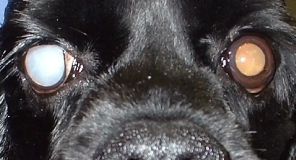
Cataracts may be inherited or acquired. Inherited cataracts can arise at any age and may vary in extent between eyes. Acquired cataracts develop secondary to diabetes (dogs), trauma, or uveitis. Despite diligent regulation of blood glucose levels, diabetic cataracts typically affect both eyes, can develop very rapidly and can result in marked inflammation within the affected eye(s) (uveitis).

Evaluation by a veterinary ophthalmologist for the rapid control of lens-induced uveitis (LIU) and/or surgical removal of the lenses (phacoemulsification) is critical. Traumatic cataracts typically develop in one eye and may not progress to affect the entire lens.

All cases of cataracts require continued monitoring for, and treatment of, LIU. Cataract removal is elective and highly successful, however like a three-legged stool, a successful surgical outcome is equally dependent upon the health of the eye, nature of the patient, and compliance of the owner.

Cataract surgery requires strict adherence to the recommended post-operative medical regimen and recheck schedule to achieve the best outcome by minimizing the development of intraocular inflammation and scarring.

Scarring that develops in an eye can result in glaucoma and/or retinal detachment, both of which can be blinding. For more information about cataracts and glaucoma, please see the related articles. (See PHH’s article on Cataracts)

Corneal Degeneration and Corneal Dystrophy

Corneal degeneration and corneal dystrophy refer to calcium or lipid deposits in the cornea(s) and can appear clinically identical. Corneal dystrophy is inherited, slowly progressive, and typically present in the central part of both corneas as white-grey crystalline deposits. The development of these deposits is not associated with prior corneal ulceration or underlying disease.

Over time, the area of deposition enlarges but at the same time, the center often becomes clear – much like an expanding doughnut. Rarely will the deposits become vision threatening.
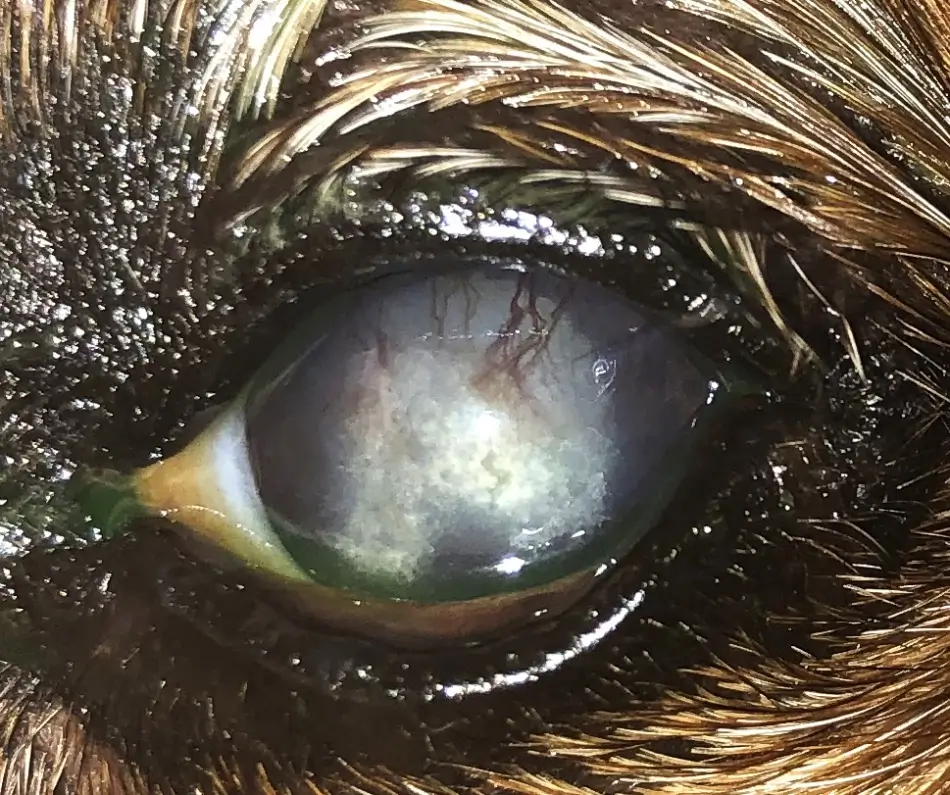
In contrast, corneal degeneration develops secondary to prior corneal injury or underlying disease (hypothyroidism, Cushing’s disease, diabetes, pregnancy, high blood cholesterol, lipid, or calcium), and may affect one cornea or both at any location.

When corneal degeneration is suspected, the underlying cause of the corneal changes can sometimes be identified through blood tests performed by your veterinarian. These blood tests will help to identify elevations in blood cholesterol, triglycerides, or calcium, and can be useful in diagnosing hypothyroidism, Cushing’s disease, and diabetes.

While uncommon, irritation of the eye by these deposits can occur, and is more often associated with calcium deposition which are often spikey and appear flakey vs the soft deposits of fat. Medication can be prescribed by your veterinarian to help blunt these sharp calcium points and promote thinning of the deposits.

Advertisement

Glaucoma

The tone or pressure of the eye is governed by the fluid within it. The pressure in the eye varies throughout the day and within a normal range. Glaucoma is defined as an elevation of intraocular pressure (IOP) that goes beyond the health of the eye.

While glaucoma is frequently blinding, early detection and treatment can prolong vision in most cases. Glaucoma is more common and more easily blinding in dogs than it is in cats.
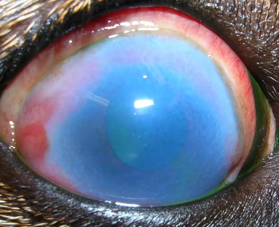
Glaucoma can be broadly classified as primary glaucoma or secondary glaucoma. Primary glaucoma is the inherited form of glaucoma that develops secondary to progressive changes that reduce fluid drainage from the eye.

Primary glaucoma will nearly always affect both eyes but not necessarily at the same time. Secondary glaucoma is acquired; caused by changes in the eye that were initiated by scarring (eg. uveitis) or mechanical obstruction within the eye (eg. space-occupying tumor, lens displacement). Secondary glaucoma typically affects one eye only.

While all dogs and cats can develop glaucoma, primary glaucoma is more common in dogs than in cats and tends to be more prevalent in certain breeds such as the American Cocker Spaniel, Basset Hound, Chow Chow, Shiba Inu, Siberian Husky, and Shar Pei. Secondary glaucoma occurs more commonly in cats than in dogs.
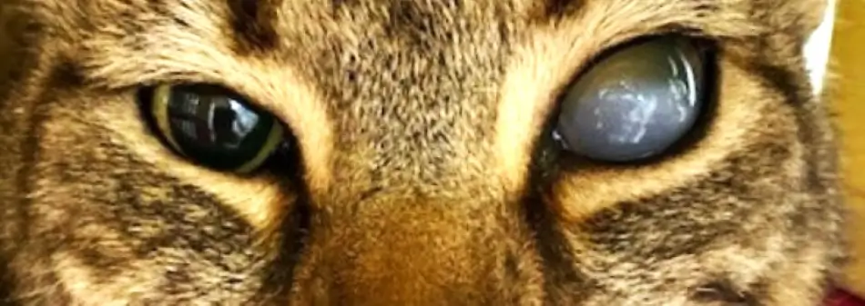
Early clinical signs associated with glaucoma (primary or secondary) can be subtle. These signs include conjunctival redness and an enlarged pupil (dilated) that does not become smaller (constricted) in response to a bright light.

Other signs that can manifest very quickly include corneal greying (some people describe this as a ‘blue’ eye), reduced vision, rubbing at the affected eye, sleeping more, eating less, or avoiding social activities.

When glaucoma is present for longer than a few days, the eye can become enlarged (buphthalmic) and in dogs is associated with blindness. Interestingly, cats can have a buphthalmic eye secondary to glaucoma but remain visual!

As discussed above, the early signs of glaucoma can be subtle and be easily missed. Because glaucoma can be rapidly blinding it is important to diagnose and start treatment early.

If you know your dog or cat to be at risk (susceptible breed, history of glaucoma in siblings or parents, history of glaucoma in the fellow eye) it is important to monitor the eyes for early signs of glaucoma.

One of the earliest indicators of elevated intraocular pressure is a dilated pupil that is poorly or not responsive to direct light stimulation. If you notice the pupil (the black center of the eye) to be dilated, you can perform the pupillary light reflex test. The pupillary light reflex test allows you to monitor the pupil’s response to a focal light source. To perform this, simply shine a bright focal light into the eye that has the dilated pupil.

Do not keep the light shining in the eye for more than a couple of seconds! You should notice the pupil become smaller very quickly in response to the light. This is called a positive direct pupillary light reflex and is a normal response to light.

A positive response does not exclude the possibility of glaucoma, and seeing your veterinarian is still warranted if other signs (described above) are noted.

When the pupil does not constrict in response to a bright focal light, this is called a negative direct pupillary light reflex. A negative or sluggish positive response warrants a call to your local veterinarian to have the IOP measured as quickly as possible.

Glaucoma is painful and very rapidly blinding. Following diagnosis of glaucoma, medication to rapidly lower eye pressure must be initiated to minimize retinal damage or blindness, and prompt referral to a veterinary ophthalmologist is recommended.

Blindness in Adult Dogs and Cats

Uncommonly, adult dogs and cats will become blind for no outwardly apparent reason. Blindness can be caused by many conditions, two of which were discussed above – glaucoma and cataracts. Blindness that occurs because of changes in the retina can occur quickly or slowly.

The two most common reasons for retinal blindness are sudden acquired retinal degeneration syndrome (SARDS) and progressive retinal atrophy (PRA). SARDS, only documented in dogs, is typified by the rapid onset (1 day to 3 weeks) of blindness with 65% of dogs reported as middle-aged to older, overweight, and female.

The cause of SARDS is unknown, but is often accompanied by increased thirst, urination, appetite, weight gain, and an initial rise in a stress hormone called cortisol. The retina of a patient affected by SARDS initially looks normal but, over several months, becomes indistinguishable from other causes of retinal atrophy.

Because the retina initially appears normal, diagnosis of SARDS requires early examination by a veterinary ophthalmologist who will be able to confirm the diagnosis of SARDS by performing a retinal function test.
This test is called electroretinography (ERG).

Unfortunately, there is no treatment for SARDS. In contrast to SARDS, PRA is inherited, slowly progressive (develops fully over 10 to 12 months), and typically develops in 3 to 5 year-old or 7 to 10 year-old age-groups of dogs. PRA can also develop in cats. By the time vision changes are noted, a patient affected by PRA has degenerative changes that include thinning of both the retina (tapetal hyperreflectivity) and retinal blood vessels (vascular attenuation).

While any breed of dog can be affected by SARDS or PRA, Dachshunds and Miniature Schnauzers are overrepresented. As with SARDS, diagnosis of PRA is confirmed via ERG testing.

Golden Retriever Uveitis

Golden Retriever Pigmentary Uveitis (GRPU) is an inherited, complex ocular condition in Golden Retrievers. The disease typically becomes evident between the ages of 4 to 7 years of age, with early signs such as intermittent tearing and redness of the conjunctival tissue, which could be mistaken for allergic conjunctivitis.

As the disease progresses, more severe symptoms develop that including aversion to light (photophobia), low intraocular pressure, darkening of the iris, and pigment deposition onto the lens.

GRU typically affects both eyes and when not treated early, can lead to chronic inflammation and intraocular scarring. Complications from these include glaucoma, cataracts, and ultimately blindness, with nearly half of affected dogs becoming blind due to glaucoma.

Early treatment focuses on controlling inflammation with topical and or oral anti-inflammatory medications and monitoring intraocular pressure.

Early detection and continuous therapy are critical, with lifelong management required to maintain vision and manage symptoms. With early intervention and continued care, the prognosis is excellent for maintenance of vision.

The prognosis becomes more guarded when the disease is detected late and intraocular scarring has already been established. Early detection emphasizes the importance of regular eye examinations to detect and manage this heritable condition effectively.

Advertisement

Conclusion

Understanding and promptly addressing eye health issues in adult dogs and cats can significantly impact their quality of life. Regular ophthalmic examinations are crucial for early detection and effective management of eye conditions.

Owners are encouraged to observe their dogs and cats at every age for any signs of eye discomfort or visual impairment and seek veterinary advice if concerns arise. Advanced treatments and surgical options are available for many conditions, which can help preserve or prolong vision.
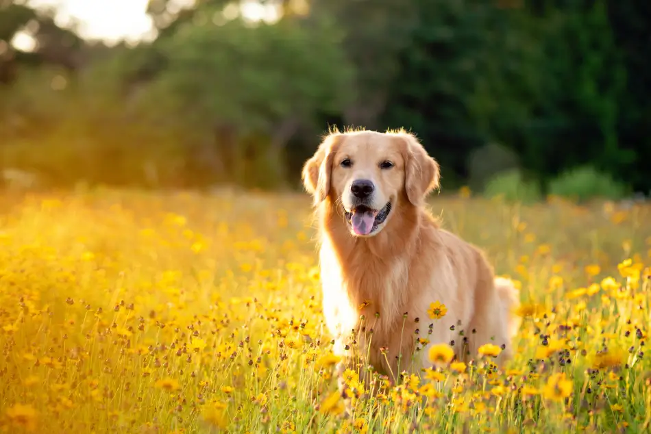
Back home, Silvia established a rigorous care regimen for Wiley. She meticulously administered the eye drops, lubricant, and thyroid medication, ensuring Wiley received his treatments on schedule. Over the ensuing weeks, she noticed remarkable improvements: Wiley’s eyes cleared, and the warmth in his gaze returned.

As his thyroid condition was brought under control, Wiley’s energy levels surged, and he even began to lose weight. Neighbors remarked on how much more playful Wiley had become, chasing squirrels in the park and greeting everyone with renewed vigor.

Thanks to Silvia’s unwavering dedication and Dr. Jensen’s expert guidance, Wiley was not just managing his conditions; he was flourishing, a testament to the enduring bond and resilience between a devoted dog and his caring owner.
Marnie Ford

By Marnie Ford PhD, DVM, Diplomate ACVO

Dr. Ford graduated from the Ontario Veterinary College in 2000 after completing a Bachelors in Zoology at the University of British Columbia and a PhD in Physiology at Monash University in Australia. Following a rotating small animal internship at the University of Minnesota in 2001, Dr. Ford achieved Diplomate status from the American College of Veterinary Ophthalmologists in 2006 after completing a 3-year residency in Comparative Ophthalmology at the University of Missouri.  After co-founding a private referral hospital, West Coast Veterinary Eye Specialists, in New Westminster (2004-2013) she opened Pacific Animal Eye Specialty services, providing mobile medical and surgical ophthalmologic care to patients in the Lower Mainland, Vancouver Island, and the Interior of BC (2013-2019).  In 2019, she returned to Australia where she worked full-time in both medical and surgical ophthalmology as well as resident training at Animal Eye Care in Melbourne Australia.   In 2023, Dr. Ford returned to Vancouver to provide locum services to ophthalmologists across Canada.

🙀

This image may be disturbing to some viewers.

Fig 1. Close up of a dog with a full white cataract in the eye on the left and no cataract in the eye on the right.
🙀

This image may be disturbing to some viewers.

Fig 3. Moist looking close up of a dog eye with milky appearance and blood vessels creeping in from the top.
🙀

This image may be disturbing to some viewers.

Fig 4. Close up of a bloodshot eye with a grey-blue bulbous cast on the cornea.
🙀

This image may be disturbing to some viewers.

Fig 5. Close up of an orange tabby cat's face with one eye normal and the other looking like a milky white orb.
🙀

This image may be disturbing to some viewers.

Fig 7. Two photos of close ups of the eye.( 7a) Normal looking brown eye, (7b) Dark brown iris with bulbous blue center.
Received from a parent or ancestor by genetic transmission
Something caused by environmental forces and not passed from parent to offspring in the genes.

Any opacification of the normally clear lens of the eye, through which light cannot pass. Can range from a tiny white speck to the entire lens being white, causing blindness in advanced cases.

Bilateral, inherited corneal disease that is usually characterized by the deposition of materials, usually lipid substances. Must be distinguished from the unilateral corneal degeneration, which occurs secondary to other corneal diseases. Can also involve the corneal epithelium and endothelium.

Deterioration of the cornea often due to aging or injury, and sometimes leading to vision impairment

Abnormally increased intraocular pressure that is incompatible with ocular health, leading to vision loss caused by damage to the optic nerve. Typically, the retina is also affected. Regular measurement of IOP (Intraocular Pressure) is crucial for early detection and management of glaucoma.

KCS, dry eye. Poor tear production and/or quality leading to a dry ocular surface that can result in discomfort, conjunctivitis, mucoid discharge, keratitis with or without corneal ulcers, corneal and/or conjunctival pigmentation, and potential blindness.

Dry eye, keratoconjunctivitis sicca. Poor tear production and/or quality leading to a dry ocular surface that can result in discomfort, conjunctivitis, mucoid discharge, keratitis with or without corneal ulcers, corneal and/or conjunctival pigmentation, and potential blindness.

GRPU. An inflammatory eye disease recognized in Golden Retrievers, characterized by pigment dispersion on the lens and iris, leading to vision loss.

Golden Retriever Pigmentary Uveitis. An inflammatory eye disease recognized in Golden Retrievers, characterized by pigment dispersion on the lens and iris, leading to vision loss.

Cherry eye. A condition commonly seen in young dogs where the third eyelid gland breaks free of its anchoring ligament attachments and prolapses, rising up to become visible as a pink-to-red mass at the leading edge of the third eyelid. Surgical repositioning of the gland is required to prevent further complications.

Chronic ulcers that are slow to heal, often occurring in the cornea

A normal age-related hardening of the lens that result in reduced focusing ability and reduced ability to see in dim light.

Not harmful in effect. In terms of a medical condition, not malignant

DM or diabetes mellitus. A metabolic disease that affects how the body uses blood sugar (glucose) as a fuel. Occurs when the pancreas doesn’t make enough insulin, and is a common cause of blinding cataracts in dogs.

Inflammation of the iris and the ciliary body (anterior uveitis) or choroid (posterior uveitis).

Inflammation of the uvea (the middle layer of the eye, including the iris, ciliary body, and choroid) triggered by immune response to lens proteins. This can occur when the lens, normally an immune-privileged site, is damaged, often due to cataracts, lens trauma, or lens aging, allowing lens proteins to leak and provoke an inflammatory reaction.

Lens-induced uveitis. Inflammation of the uvea (the middle layer of the eye, including the iris, ciliary body, and choroid) triggered by immune response to lens proteins. This can occur when the lens, normally an immune-privileged site, is damaged, often due to cataracts, lens trauma, or lens aging, allowing lens proteins to leak and provoke an inflammatory reaction.

The surgical procedure of breaking the lens up with ultrasound and aspiration fragments.

A common hormonal disorder in dogs in which the thyroid gland fails to produce enough thyroid hormone. This deficiency can lead to a variety of signs including lethargy, weight gain, hair loss, and skin problems. It is particularly prevalent in medium to large breeds, and requires lifelong treatment with thyroid hormone replacement therapy.

A hormonal condition where the adrenal glands produce an excessive amount of cortisol, either secondary to an adrenal or pituitary tumor. This hormonal imbalance can lead to various signs in dogs, such as increased thirst and urination, increased appetite, hair loss, and a pot-bellied appearance. It's often seen in middle-aged to older dogs and requires careful diagnosis and lifetime management.

IOP. The fluid pressure inside the eye, critical for maintaining the shape of the eye and preserving healthy vision.

Intraocular pressure. The fluid pressure inside the eye, critical for maintaining the shape of the eye and preserving healthy vision.

A type of glaucoma that occurs without a known secondary cause, often associated with increased intraocular pressure leading to optic nerve damage.
Glaucoma that develops as a result of another eye condition or disease.

The black opening in the center of the iris that, by changing shape, regulates the amount of light that enters the eye.

For the eye, dilated refers to pupils that are larger than normal.
An eye condition where one or both eyeballs are larger than normal. Often associated with increased intraocular pressure, a condition known as glaucoma.
PLR. A reflex that controls the diameter of the pupil in response to the intensity of light. An objective parameter in assessing the function of the retina, optic nerve, and oculomotor nerve.
Constriction of the pupil in response to light shone directly into the eye.

SARDS. A disease in dogs causing sudden blindness due to rapid degeneration of the retina. The exact cause is unknown, and sadly, there is no known effective treatment or cure for SARDS as of now.

Sudden acquired retinal degeneration syndrome. A disease in dogs causing sudden blindness due to rapid degeneration of the retina. The exact cause is unknown, and sadly, there is no known effective treatment or cure for SARDS as of now.

PRA. An inherited degenerative blinding disease of the retina, occurring commonly in dogs and rarely in cats. Blindness occurs slowly and progressively due to death of retinal cells.

Progressive Retinal Atrophy. An inherited degenerative blinding disease of the retina, occurring commonly in dogs and rarely in cats. Blindness occurs slowly and progressively due to death of retinal cells.

Loss of tissue structure after normal development; applied mainly to degenerative hereditary diseases of mature animals.

Recording of retinal electrical potentials generated by a rapid change in illumination. Divided into flash and pattern types.

Electroretinogram.

The innermost, light-sensitive layer of tissue in the back of the eye that contains the light sensitive rods and cones.

Increased reflection of light from the tapetum, a reflective layer behind the retina, often seen in retinal degenerative conditions.
Pertaining to the retina, the innermost, light-sensitive layer of tissue in the back of the eye that contains the light sensitive rods and cones.
Reduction in the diameter or density of blood vessels, often observed in retinal imaging.

Abnormal light sensitivity. Often indicating eye disease or inflammation, photophobia can be a symptom of conditions like uveitis, corneal ulceration, or cataracts.

The thin coloured part of the eye surrounding the pupil that controls the amount of light entering the eye by adjusting pupil size. The iris contains smooth muscle, pigment cells and blood vessels.

Located behind the iris, the lens is a curved, transparent, biconvex structure suspended in the eye that is flexible to bend light and focus it on the retina to help you see images clearly.

A condition or situation that persists and often recurs over a long period of time.
A biological response to harmful stimuli, such as pathogens or irritants, characterized by redness, swelling, heat, and pain

Advertisement

🙀

This image may be disturbing to some viewers.

Fig 1. Close up of a dog with a full white cataract in the eye on the left and no cataract in the eye on the right.
🙀

This image may be disturbing to some viewers.

Fig 3. Moist looking close up of a dog eye with milky appearance and blood vessels creeping in from the top.
🙀

This image may be disturbing to some viewers.

Fig 4. Close up of a bloodshot eye with a grey-blue bulbous cast on the cornea.
🙀

This image may be disturbing to some viewers.

Fig 5. Close up of an orange tabby cat's face with one eye normal and the other looking like a milky white orb.
🙀

This image may be disturbing to some viewers.

Fig 7. Two photos of close ups of the eye.( 7a) Normal looking brown eye, (7b) Dark brown iris with bulbous blue center.
Received from a parent or ancestor by genetic transmission
Something caused by environmental forces and not passed from parent to offspring in the genes.

Any opacification of the normally clear lens of the eye, through which light cannot pass. Can range from a tiny white speck to the entire lens being white, causing blindness in advanced cases.

Bilateral, inherited corneal disease that is usually characterized by the deposition of materials, usually lipid substances. Must be distinguished from the unilateral corneal degeneration, which occurs secondary to other corneal diseases. Can also involve the corneal epithelium and endothelium.

Deterioration of the cornea often due to aging or injury, and sometimes leading to vision impairment

Abnormally increased intraocular pressure that is incompatible with ocular health, leading to vision loss caused by damage to the optic nerve. Typically, the retina is also affected. Regular measurement of IOP (Intraocular Pressure) is crucial for early detection and management of glaucoma.

KCS, dry eye. Poor tear production and/or quality leading to a dry ocular surface that can result in discomfort, conjunctivitis, mucoid discharge, keratitis with or without corneal ulcers, corneal and/or conjunctival pigmentation, and potential blindness.

Dry eye, keratoconjunctivitis sicca. Poor tear production and/or quality leading to a dry ocular surface that can result in discomfort, conjunctivitis, mucoid discharge, keratitis with or without corneal ulcers, corneal and/or conjunctival pigmentation, and potential blindness.

GRPU. An inflammatory eye disease recognized in Golden Retrievers, characterized by pigment dispersion on the lens and iris, leading to vision loss.

Golden Retriever Pigmentary Uveitis. An inflammatory eye disease recognized in Golden Retrievers, characterized by pigment dispersion on the lens and iris, leading to vision loss.

Cherry eye. A condition commonly seen in young dogs where the third eyelid gland breaks free of its anchoring ligament attachments and prolapses, rising up to become visible as a pink-to-red mass at the leading edge of the third eyelid. Surgical repositioning of the gland is required to prevent further complications.

Chronic ulcers that are slow to heal, often occurring in the cornea

A normal age-related hardening of the lens that result in reduced focusing ability and reduced ability to see in dim light.

Not harmful in effect. In terms of a medical condition, not malignant

DM or diabetes mellitus. A metabolic disease that affects how the body uses blood sugar (glucose) as a fuel. Occurs when the pancreas doesn’t make enough insulin, and is a common cause of blinding cataracts in dogs.

Inflammation of the iris and the ciliary body (anterior uveitis) or choroid (posterior uveitis).

Inflammation of the uvea (the middle layer of the eye, including the iris, ciliary body, and choroid) triggered by immune response to lens proteins. This can occur when the lens, normally an immune-privileged site, is damaged, often due to cataracts, lens trauma, or lens aging, allowing lens proteins to leak and provoke an inflammatory reaction.

Lens-induced uveitis. Inflammation of the uvea (the middle layer of the eye, including the iris, ciliary body, and choroid) triggered by immune response to lens proteins. This can occur when the lens, normally an immune-privileged site, is damaged, often due to cataracts, lens trauma, or lens aging, allowing lens proteins to leak and provoke an inflammatory reaction.

The surgical procedure of breaking the lens up with ultrasound and aspiration fragments.

A common hormonal disorder in dogs in which the thyroid gland fails to produce enough thyroid hormone. This deficiency can lead to a variety of signs including lethargy, weight gain, hair loss, and skin problems. It is particularly prevalent in medium to large breeds, and requires lifelong treatment with thyroid hormone replacement therapy.

A hormonal condition where the adrenal glands produce an excessive amount of cortisol, either secondary to an adrenal or pituitary tumor. This hormonal imbalance can lead to various signs in dogs, such as increased thirst and urination, increased appetite, hair loss, and a pot-bellied appearance. It's often seen in middle-aged to older dogs and requires careful diagnosis and lifetime management.

IOP. The fluid pressure inside the eye, critical for maintaining the shape of the eye and preserving healthy vision.

Intraocular pressure. The fluid pressure inside the eye, critical for maintaining the shape of the eye and preserving healthy vision.

A type of glaucoma that occurs without a known secondary cause, often associated with increased intraocular pressure leading to optic nerve damage.
Glaucoma that develops as a result of another eye condition or disease.

The black opening in the center of the iris that, by changing shape, regulates the amount of light that enters the eye.

For the eye, dilated refers to pupils that are larger than normal.
An eye condition where one or both eyeballs are larger than normal. Often associated with increased intraocular pressure, a condition known as glaucoma.
PLR. A reflex that controls the diameter of the pupil in response to the intensity of light. An objective parameter in assessing the function of the retina, optic nerve, and oculomotor nerve.
Constriction of the pupil in response to light shone directly into the eye.

SARDS. A disease in dogs causing sudden blindness due to rapid degeneration of the retina. The exact cause is unknown, and sadly, there is no known effective treatment or cure for SARDS as of now.

Sudden acquired retinal degeneration syndrome. A disease in dogs causing sudden blindness due to rapid degeneration of the retina. The exact cause is unknown, and sadly, there is no known effective treatment or cure for SARDS as of now.

PRA. An inherited degenerative blinding disease of the retina, occurring commonly in dogs and rarely in cats. Blindness occurs slowly and progressively due to death of retinal cells.

Progressive Retinal Atrophy. An inherited degenerative blinding disease of the retina, occurring commonly in dogs and rarely in cats. Blindness occurs slowly and progressively due to death of retinal cells.

Loss of tissue structure after normal development; applied mainly to degenerative hereditary diseases of mature animals.

Recording of retinal electrical potentials generated by a rapid change in illumination. Divided into flash and pattern types.

Electroretinogram.

The innermost, light-sensitive layer of tissue in the back of the eye that contains the light sensitive rods and cones.

Increased reflection of light from the tapetum, a reflective layer behind the retina, often seen in retinal degenerative conditions.
Pertaining to the retina, the innermost, light-sensitive layer of tissue in the back of the eye that contains the light sensitive rods and cones.
Reduction in the diameter or density of blood vessels, often observed in retinal imaging.

Abnormal light sensitivity. Often indicating eye disease or inflammation, photophobia can be a symptom of conditions like uveitis, corneal ulceration, or cataracts.

The thin coloured part of the eye surrounding the pupil that controls the amount of light entering the eye by adjusting pupil size. The iris contains smooth muscle, pigment cells and blood vessels.

Located behind the iris, the lens is a curved, transparent, biconvex structure suspended in the eye that is flexible to bend light and focus it on the retina to help you see images clearly.

A condition or situation that persists and often recurs over a long period of time.
A biological response to harmful stimuli, such as pathogens or irritants, characterized by redness, swelling, heat, and pain

Already have an account?

Two ways to read this article:

By continuing, you agree to the Privacy Policy, Terms of Use