 Pet News


Summary
This article explores the unique anatomical features and associated health risks of brachycephalic breeds—dogs with notably shortened upper jaws and noses, such as Pugs, French Bulldogs, and Bulldogs. These breeds are characterized by a brachycephalic ocular syndrome, which includes a shallow orbital structure, large eyelid openings, and protruding eyes, leading to significant ocular health issues.
The discussion highlights how these physical traits increase the risk for corneal ulcers, proptosis (eye displacement), and other serious eye conditions. The text delves into the anatomical reasons behind these issues, such as the shallow orbit and large eyelid openings that fail to adequately protect or cover the eye.
It also covers potential solutions and management strategies, including surgical interventions and the application of corneal lubricants, to mitigate these risks. The overall aim is to raise awareness of the health challenges faced by brachycephalic breeds and to promote responsible breeding and care practices to ensure the wellbeing of these dogs.
Pudge, a brownish-black Pug puppy, captured hearts with his adorable round eyes and wrinkled face. His owner, Emily, fell in love with him instantly, unaware of the unique challenges his breed faced.
As Pudge grew, so did concerns about his ocular health. Despite his cuteness, Pudge’s shallow orbits and large eyelid openings put him at risk for serious eye conditions. Emily noticed his eyes watering excessively and occasional squinting, prompting a visit to the veterinarian.
The vet diagnosed Pudge with corneal ulcers, a common issue among brachycephalic breeds like Pugs. Emily was devastated but determined to help her furry friend. Following the vet’s advice, she diligently applied corneal lubricants to ease Pudge’s discomfort and minimize the risk of further complications.
Despite his struggles, Pudge remained cheerful and resilient. Emily marveled at his unwavering spirit, finding strength in his playful antics and unwavering affection. She made sure Pudge received the best care possible, including regular check-ups and measures to protect his delicate eyes.
Through Pudge’s journey, Emily became an advocate for responsible breeding and proactive healthcare in brachycephalic breeds. She shared Pudge’s story to raise awareness about the importance of prioritizing health over appearance, ensuring that other adorable pups like Pudge could live happy, healthy lives.
In the end, Pudge’s story wasn’t just about overcoming health challenges—it was a testament to the unbreakable bond between a devoted owner and her beloved pup.
Introduction
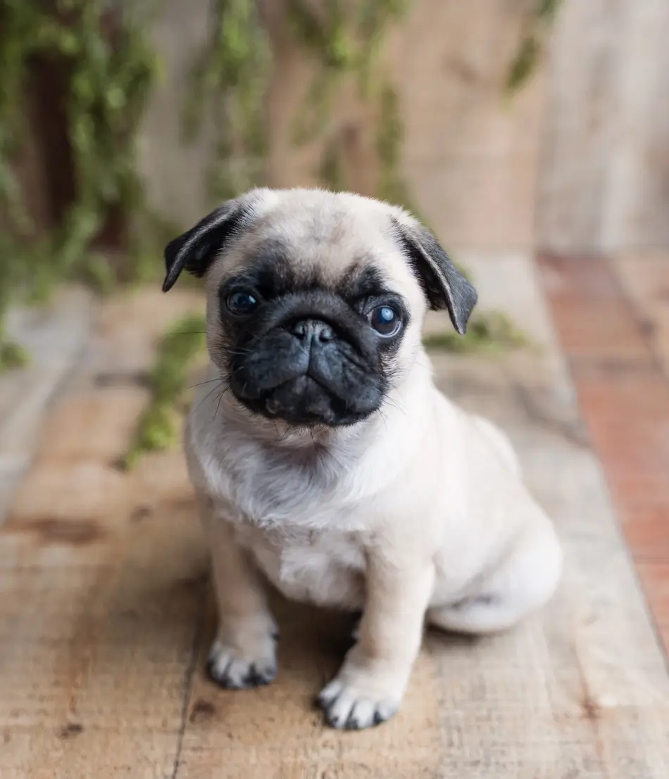
Animals who have greatly shortened upper jaws and noses are called brachycephalic. Some of the more common brachycephalic breeds include Pugs, Pekingese, Boston Terriers, French Bulldogs, and Bulldogs. This head shape also commonly includes exophthalmos (abnormal protrusion of the eyes), exaggerated eyelid openings (macropalpebral fissure), and lagophthalmia (inability to completely close the eyelids). Collectively known as macropalpebral ocular syndrome (or brachycephalic ocular syndrome) these traits create large, “baby-like,” round eyes that are frequently the impetus that attracts people to these breeds.
Advertisement
The “Cute” Factor
Sadly, the “cute” factor poses unique risks to long-term ocular health and vision. Through continued selection of these exaggerated conformational traits that include shallow orbits, large eyelid openings, reduced corneal sensitivity, and low tear production, ocular problems are overrepresented. Brachycephalic breeds have been reported to be 8 to 20 times more likely to develop a corneal ulcer compared to non-brachycephalic breeds, and the ulcers they suffer from are often deeper than those in non-brachycephalic breeds. The reasons, solutions, and mitigation options for this are outlined below.
Skull Shapes

The difference in skull shape between brachycephalic breeds (e.g., Pug) and dolichocephalic breeds (e.g., Greyhound) is striking. The severely shortened skull and muzzle of the brachycephalic head are inherited. The soft tissues of the head, however, are not similarly reduced in size, and herein lies the source of most ocular problems. In all breeds of dog, the bony orbit is made up by seven bones and contains access routes for vessels, nerves, muscles that control eye movement, and glands, as well as containing connective tissue and fat. Unlike the orbit of the dolichocephalic or long nosed breeds, which surrounds and protects the eye, the orbit of brachycephalic head shape is much shallower—like a pie plate rather than a bowl.

Photo credit: M Sanders
With the normal extraocular structures listed above and the shallowness of the space, it is easy to see how precarious the globe’s position within orbit is! This anatomic shortcoming can be easily felt by touching the top of the bony orbit (the orbital rim) where it sits behind the eyeball and unable to provide any protection to the eye. In other words, looking at the profile of the dog head, the curve of the eyeball extends well beyond the rim of the orbit.
The Problem with Very large Eyelid Openings
Very large eyelid openings contribute to both incomplete eyelid closure and coverage of the eye. These, coupled with a shallow orbit, increase the risk for proptosis (displacement of the eye), following minor forms of trauma that may involve sudden forces applied to the head and orbit, stretching of the facial skin (e.g., scruffing the dog’s neck, or during a dogfight) or even increased neck pressure (e.g., from pulling on a leash).
Following the forward movement of the globe through the eyelid openings, the eyelids quickly close behind the globe, thus trapping it in this forward and very painful position. Spontaneous return to the loosely defined “normal” position is hindered by bleeding behind the eye, swelling, possible tearing of the eye muscles, and the presence of the eyelid margins behind the globe. Without rapid manual replacement of the globe, tension on the optic nerve may result in permanent vision impairment, and rapid drying of the exposed cornea can result in corneal ulceration.
This is not the case in dolichocephalic breeds, for which significant force is required to dislodge the eye, and vision can rarely be saved. To test the ability for complete eyelid closure, the corners of the eyelids closest to the nose, and closest to the ear, can be lightly touched to stimulate the blink response. Similarly, check to see if your dog is fully closing his/her lids when fully asleep. An incomplete ability to blink fully not only increases the risk for displacement of the eye, as outlined above, but also increases the risk for direct damage by external objects and indirect damage by reduced spread of the tear film—a major factor in corneal drying, ulceration, and pigment deposition.
To help reduce the potential consequences of incomplete eyelid closure, frequent application of a corneal lubricant is strongly recommended. However, surgical narrowing of the inner and sometimes outer corners of the eyelids can be performed for more effective protection. This surgery is called a medial canthoplasty and/or lateral canthoplasty.
Corneal Pigmentation

Corneal pigmentation is very common in brachycephalic breeds. The incidence reported in Pugs varies from 70.0 to 87.8 per cent. Not unlike skin chronically irritated by rubbing, corneal pigmentation forms as a protective mechanism, and in brachycephalic breeds is frequently caused by some degree of medial lower lid entropion associated with rounded eyelid openings. See article on Entropion.
Unfortunately, this protective mechanism is not conducive to unobstructed vision. When noted during wellness examinations, corneal pigment can signal the presence of conformational (entropion, over exposure) irritation that may be mitigated surgically or medically as outlined above. Other sources of corneal irritation associated with the eyelids include caruncular hairs, distichia, trichiasis from exaggerated nasal folds, and ectopic cilia. The pigment can be increased by corneal blood vessels and explains why corneal pigment is found to be worse after a corneal ulcer. See article on Corneal Ulcers.
Ectopic cilia can be present in any breed of dog, but in my experience, seem to be more prevalent in the Pug. Unless these sources of irritation are eliminated or reduced with surgery (especially ectopic cilia) or application of a thick corneal lubricant, pigment in some patients, especially Pugs, can cause blindness.
Advertisement
Excessive Tearing
Excessive tearing (epiphora) is a common finding in brachycephalic dogs. Normally tears drain through a small opening (punctum) on the inside of each eyelid near the bridge of the nose, via the tear duct into the nose. In brachycephalic dogs, this drainage is often disrupted by kinking of the tear duct due to the excessive skin folds that results from the short facial bones and normal soft tissue length, and/or blocking of the tear puncta by the large, protruding eyes. Excessive tearing which causes tears to overflow onto the face results in tear-staining and dermatitis (“tear burn”). See article on Itchiness (Pruritus) in Dogs. For more information, please see the Related Articles to this article.
How Skull Shape Affects the Cornea
The short nose of the brachycephalic skull shape directly affects the safety of the cornea in two ways. Unlike a longer nose, which acts as a bumper between the cornea and external objects, the short nose of a brachycephalic breed puts the cornea under constant threat of direct trauma by whatever the dog is sniffing. Likewise, actions such as rubbing the mouth or scratching the ears or shaking the head (if ears are long and hairy) create a daily risk of damage to the globe that is compounded by incomplete lid closure. The length of the nose is also associated with corneal sensitivity.
Reduction in Response to Pain
Researchers have demonstrated an additional vulnerability of the eyes of brachycephalic breeds, which is that they have a smaller number of pain nerves in the cornea than dogs with longer noses. Reduced corneal sensitivity, which invariably ensues, reduces the dog’s response to pain (e.g., squinting, rubbing, tearing) following irritation or injury, which in turn delays owner awareness and makes owners less likely to seek treatment. Unfortunately, it is often a deep defect or corneal greying that is seen that treatment is initiated. It is for this reason that ulcers in brachycephalic dogs are initially diagnosed by a veterinarian at a more advanced stage (deeper, more infected, ruptured) than those of longer-nosed breeds. You must never ignore any corneal colour change, increased or persistent squinting, or tearing in any breed, but especially the brachycephalic breeds. To minimize direct trauma to the eyes, goggles can be worn by your dog.
The Trifecta
The brachycephalic dog has the trifecta of eye problems: shallow orbit, overly large eyelids, and a short nose. One might think that these “design faults” were enough to contend with, but . . . sadly, no. The consequences of each are compounded by the presence of low tear production and/or low tear film quality (keratoconjunctivitis sicca, dry eye). See article on Dry Eye, and rarely does one problem develop independent of at least one other factor. Tear production has been demonstrated to be lower in brachycephalic than in non-brachycephalic dogs, and this is thought to be linked to the reduced corneal sensitivity. The consequences of keratoconjunctivitis sicca include blood vessel growth into the cornea, pigmentation, scarring, ulceration, and possible blindness. Tear production should be measured via a Schirmer tear test in every brachycephalic patient at every examination, and tear film quality assessed.
Advertisement
Conclusion
Despite their many design flaws, we love our brachycephalic breeds for their delightful personalities, inquisitiveness, and size. However, brachycephalic eye health is precarious and the normalization of extreme conformational traits that cause discomfort and threaten vision should be unacceptable. Through education and awareness of how specific breed traits can affect health, selection of puppies without these extreme traits, and early care of those with them, can go a long way towards providing a pain-free and healthy life for these delightful companions.

In the end, Pudge’s story wasn’t just about overcoming health challenges—it was a testament to the unbreakable bond between a devoted owner and her beloved pup.
Through Pudge’s journey, Emily became an advocate for responsible breeding and proactive healthcare in brachycephalic breeds. She shared Pudge’s story to raise awareness about the importance of prioritizing health over appearance, ensuring that other adorable pups like Pudge could live happy, healthy lives.
In the end, Pudge’s story wasn’t just about overcoming health challenges—it was a testament to the unbreakable bond between a devoted owner and her beloved pup.
By Marnie Ford PhD, DVM, Diplomate ACVO
Dr. Ford graduated from the Ontario Veterinary College in 2000 after completing a Bachelors in Zoology at the University of British Columbia and a PhD in Physiology at Monash University in Australia. Following a rotating small animal internship at the University of Minnesota in 2001, Dr. Ford achieved Diplomate status from the American College of Veterinary Ophthalmologists in 2006 after completing a 3-year residency in Comparative Ophthalmology at the University of Missouri. After co-founding a private referral hospital, West Coast Veterinary Eye Specialists, in New Westminster (2004-2013) she opened Pacific Animal Eye Specialty services, providing mobile medical and surgical ophthalmologic care to patients in the Lower Mainland, Vancouver Island, and the Interior of BC (2013-2019). In 2019, she returned to Australia where she worked full-time in both medical and surgical ophthalmology as well as resident training at Animal Eye Care in Melbourne Australia. In 2023, Dr. Ford returned to Vancouver to provide locum services to ophthalmologists across Canada.