 Pet News

Summary
A feline corneal sequestrum is a chronic medical condition in cats where a portion of the cornea (the transparent outer layer of the eye) dies and becomes “mummified”. It is commonly seen as a brown-to-black spot on the cornea and can cause discomfort, severe corneal inflammation, and vision problems for the affected cat.
The condition requires surgical removal accompanied by lifetime lubrication, to alleviate pain and prevent further complications. Breeds of cats with prominent eyes are especially at risk for developing corneal sequestra.
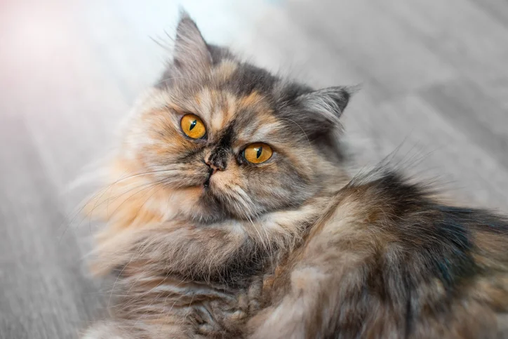
I have had so many Persian cat patients with corneal sequestra! They all needed lots of help and the owners needed lots of guidance. Read on, to learn more about this mysterious condition that afflicts primarily bug-eyed cats such as Persians, Exotic Shorthairs, and Burmese.

Maggie thinks that her three year old Persian cat, “Opalina”, has the most beautiful eyes in the world. Huge and mysterious, they don’t miss a thing. Maggie’s one regret is that Opalina’s striking eyes are always runny, with dark tear stains that make her look a bit like a raccoon.
Opalina was healthy except that about once a year, she would have a cold, making her eyes run more and be a little squinty for a month or so, especially the left eye. Maggie has a very busy job that keeps her away from home for long hours, but when she is home Opalina’s eyes are always watching her, unblinking and constant—especially at breakfast and dinnertime.
One day, at kitty dinner time, Maggie noticed that the left eye looked a bit odd and the cat was slightly squinting that eye. Looking closer, she saw that there appeared to be a dark smudge on the surface but decided she must be wrong and that it was really the normal black pupil.
Over the next few months, Opalina squinted just a little bit at times and the tear staining on the left side worsened. Then one morning Opalina didn’t show up for breakfast and Maggie found her hiding under the bed.
Maggie was horrified to see that the left eye was nearly swollen shut and the surface was bluish-white except for a piece of “something”, which showed up black against the cloudy cornea.
Maggie called her veterinarian’s office but could not get an appointment for 2 weeks. She ended up waiting until 5:00 pm when the local emergency clinic opened. The emergency veterinarian thought the eye looked like it had been scratched, which Maggie knew was impossible.
The veterinarian recommended referral ASAP to a veterinary ophthalmologist, and in the meantime prescribed oral pain medication, an E collar, and a topical antibiotic eye drop. Thanks to the emergency veterinarian, Opalina was “fast-tracked” and seen by me as an emergency work-in patient one day later.
Introduction
Feline corneal sequestration (singular: sequestrum. plural: sequestra) is a dramatic, complicated and unusual corneal disease unique to domestic cats, and refers to a bizarre change of color of the cornea due to the development of an opaque, dark brown to black plaque.
This plaque is actually a dead piece of cornea firmly embedded, or “sequestered” within surrounding living corneal tissue. Another term for this condition is “corneal mummification”. It is a mystery of nature as to how living corneal tissue can tolerate a patch of dead corneal tissue tightly joined to neighboring living corneal cells.
Usually only one eye is affected. but some cats (especially Persian cats) will eventually have both eyes affected, although it is quite rare to have both eyes affected at the same time. Advanced corneal sequestra are often painful and can lead to rupture and surgical removal of the eye if not adequately treated. Treatment often requires surgery to remove the sequestrum, but many cats can be effectively managed medically.
It is important to know that corneal sequestra are rare enough that most general practitioner veterinarians do not recognize the condition in an affected animal, even in advanced cases.
Corneal sequestra are difficult to both diagnose and treat by general practitioner veterinarians, and all affected cats (or presumed to be affected cats) really should be referred to a veterinary ophthalmologist for diagnosis, outline of treatment options, and care.
If your cat has a brownish to blackish discoloration of one or both eyes, referral to a veterinary ophthalmologist is recommended, as treatment of corneal sequestra is best managed by an ophthalmologist.
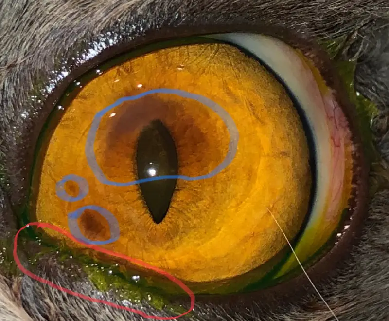
Corneal sequestra are usually oval to round, can be quite small to quite large, and they can eventually extend deeply into the corneal tissue, even involving the full thickness of the cornea in advanced cases. Early in the disease, affected corneas start out with mild discoloration, from lightest amber to a “bronzed” appearance that progresses to brown and then to black (brown discoloration of a tissue is termed “brunescence”).
Early sequestra often look like a faint dusky shadow on the cornea, so they are typically not noticed by the owner or a general practitioner DVM until later when they are larger and darker.
Advanced sequestra often have the appearance of a shiny raised piece of black patent leather on the surface of the eye. Even then, the condition can be missed because the owner often assumes that the dark patch on the cornea is the normal black pupil!

A corneal sequestrum can be accompanied by an overlying corneal ulcer or with no ulcer present. The outermost layer of the normal cornea is the thin epithelium, which is similar to the epithelium in the surface of skin.
If the epithelium has a patch missing, it is a type of ulcer called a corneal erosion and is painful due to exposed nerve endings. If a patch of deeper corneal tissue (stroma) is also missing, this type of ulcer is a corneal stromal ulcer and is also painful.
Erosions are sometimes termed ulcers. Just as skin wounds can be superficial or deeper, so can corneal wounds be superficial (erosion) or deeper (stromal ulcer) or very deep (deep stromal ulcer).

B. Diagram of very early corneal sequestrum and overlying corneal erosion. Faint "bronzed" corneal stroma is present.
C. Diagram of more advanced corneal sequestrum with overlying corneal erosion. Dark brown corneal stroma is present.
Diagrams courtesy of Marnie Ford DVM
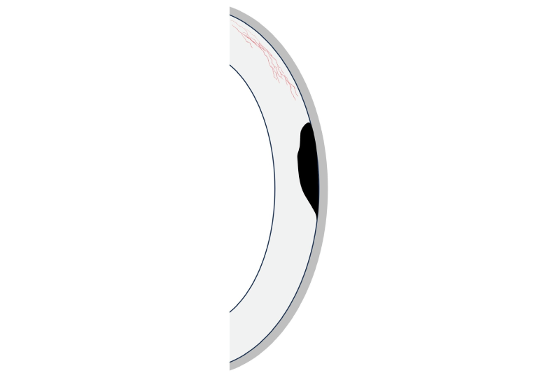

B. Diagram of "quietly" extruding advanced corneal sequestrum. There is increased corneal vascularization, that has reached the sequestrum, and the sequestrum is starting to "lip" and lift off the cornea on one edge.
C. Diagram of extruding full-thickness corneal sequestrum with melting cornea (green) surrounding it. The sequestrum will fall off of the eye and the eye will then rupture.
Courtesy of Marnie Ford DVM

Courtesy of Marnie Ford DVM
Courtesy of Marnie Ford DVM

Courtesy of Marnie Ford DVM

Courtesy of Marnie Ford DVM
Cause and Risk Factors for Corneal Sequestration
The exact cause of feline sequestra is unknown. Genetics and head conformation may be involved since there is a definite breed predisposition.
Corneal sequestration can occur in cats of all ages and breeds, with brachycephalic breeds most at risk. This breed predilection might be due to genetics, and/or head conformation and prominence of the eyes and the conformation of the eyelids and (if present) nasal folds.
The breed with the highest frequency of occurrence is the Persian breed. Other at-risk breeds include Siamese, Himalayan, Burmese, and Bombay.
There is often a corneal ulcer overlying the sequestrum, which can be superficial (erosion) or deep (stromal ulcer). An ulcer is a risk factor for a sequestrum to form. The degree of pain depends on how much living corneal tissue is involved.
For example, if an ulcer only exposed the underlying sequestrum, there may not be any pain because the sequestrum is dead tissue with no nerve endings.
An ulcer is classically diagnosed by the application of a green dye called fluorescein dye, which sticks to the underlying living corneal tissue and stains it green. However, when the underlying tissue is dead tissue in a sequestrum, it won’t stain well, so an ulcer can be easily missed by a general practitioner veterinarian.
Other risk factors include:
- Trauma
- Chronic nasal fold trichiasis (eyelid hair rubbing on the cornea). This is especially common in Persian cats, with hair on the nasal fold rubbing on the cornea.
- Chronic entropion (rolling-in of the eyelid, causing hair to rub on the cornea). This is especially common in Persian cats, in the inside (medial) corners of the lower eyelids.
- Feline Herpesvirus (FHV-1) infection
- Dry Eye (not enough tears, and/or poor quality tears)
- Poor blink (lagophthalmos). Brachycephalic cats do not blink well compared with normal cats.
- Minor surgery (grid keratotomy) performed on eyes with chronic superficial corneal ulcers. This surgery is typically very successful in dogs, but should not be performed in cats with chronic ulcers.
- If the other eye had a sequestrum, then the remaining eye is at high risk to also develop one (or more!) at any time in the future– especially brachycephalic cats.
Keep in mind that brachycephalic cats have prominent eyes that are inherently more prone to trauma, nasal fold trichiasis, medial entropion, and to poor blink. Persian cats tend to develop the darkest sequestra, because this breed is prone to naturally having dark reddish-brown tears.
These tears are often mistaken for dried blood, but are normal for many cats, including some non-Persian cats. The cause of the tear discoloration is unknown, but the pigment can end up staining the sequestra, making them darker.
Clinical Signs of Corneal Sequestration
A thorough ophthalmic examination, preferably by a veterinary ophthalmologist, is essential to uncover any underlying problems that might also need to be addressed. Sequestra can be painful. The clinical signs of ocular pain may be subtle or severe and include:
- Depression (lethargy, sleeping excessively, reduced appetite)
- Squinting
- Increased Tearing
- Resistance to petting around the head
Other clinical signs which may be present include:
- Cloudiness
- Discharge (watery or mucoid; clear, white, gray, reddish-brown, green/yellow, or blood-tinged)
- Brown to black corneal discoloration
- Corneal ulcer or erosion
- An elevation or “bump” on the surface of the cornea (this is an extruding sequestrum, or if the eye has ruptured, it is the rupture site– or both).
- A “crater” on the surface of the cornea (if the sequestrum suddenly extruded and fell off of the cornea)
- Redness of the conjunctiva and or cornea.
- Blood on the surface of the eye (if the cornea ruptured)
- Elevation of the third eyelid (due to pain and guarding of the eye)
Advertisement
Diagnosis
Diagnosis is by visualization of the darkened, discolored cornea. Examination of the cornea is best performed by an ophthalmologist with high magnification ( biomicroscopy) to determine the severity of the sequestrum.
Other tests that are usually performed include fluorescein dye application to determine if an ulcer is present, and also a Schirmer tear test to measure tear volume. Sometimes a tear film breakup time test or a Rose Bengal dye test will be performed to determine the tear quality.
Corneal sequestra are relatively rarely encountered by general practitioners, but if a sequestrum is suspected, most general practitioners will refer to these patients. Diagnosis often does not occur until the cat is referred to a veterinary ophthalmologist.
Clinical Course of Corneal Sequestra
Sequestra can remain for months to years, appearing to be “no big deal”, but can eventually and silently progress until there is a sudden crisis.
The dead corneal tissue exists immediately next to living corneal tissue for months to years, and thus can act as a foreign body– eventually “waking up” the immune system to the fact that this dead tissue is present, causing the immune system to attack the sequestrum to try to get rid of it.
During this attack, blood vessels carrying inflammatory cells grow into the cornea. In some cases, no blood vessels invade the cornea, but neighboring healthy corneal tissue is still attacked and can suddenly break down and melt.
A sequestrum can break free of the surrounding living cornea and extrude (slough) from the eye’s surface. Extrusion is unpredictable! It can be very slow or very fast.
If it is slow, then luck may be with the cat and the sequestrum slowly and quietly develops a ‘lipped edge’ and then lifts off of the underlying healthy cornea and eventually falls off, somewhat like a hangnail forming.
This quiet, slow process is usually not painful; remember that the sequestrum itself is dead tissue, so it does not have any nerve endings.
However, if extrusion is rapid, it is because the immune attack was sudden and intense, causing neighboring corneal tissue to melt – sometimes in a matter of hours.
This is usually quite painful. In this case, as the sequestrum extrudes, it can leave a big crater (deep corneal ulcer, or even a full-thickness hole in the cornea) and the eye can rupture– especially if the sequestrum involves the full thickness of the cornea.
If rupture occurs, the eye needs surgery– usually it cannot be saved and needs surgical removal ( enucleation), but sometimes it can be saved with major surgery to place a tissue graft.
It is impossible for an ophthalmologist to determine whether an early or mid-stage sequestrum will “behave itself” and not later extrude quickly via rapid melting and the eye rupture.
It is not advised to let Mother Nature take its course and let the sequestrum extrude on its own, as the process is so unpredictable: in many cases, extrusion starts out quietly with minimal immune attack and inflammation, but the immune system can suddenly “wake up” and attack the cornea at any time– literally overnight— and the cornea can rapidly melt and rupture. Having a corneal sequestrum is like having a ticking “eye bomb” which may or may not go off at any time.
Besides the risk of a sequestrum later extruding suddenly, violently, and painfully (leading to possible surgical removal of the eye), another risk to choosing Mother Nature’s course is that even if the cornea does heal, it could heal with much scarring (reducing vision), and/or it is quite possible that the eye will develop more sequestra later– especially in Persian cats.
If surgery is not elected, then daily lifetime supportive treatment (especially in a brachycephalic cat) is needed to protect BOTH eyes— remember that if one eye has a sequestrum, the other eye is at high risk to also develop a sequestrum. So taking preventative measures are important!
Advertisement
Treatment
Since sequestra are painful, can remain for months to years, can seriously affect vision, and can lead to eye rupture, early surgical removal of the sequestrum is the treatment of choice.
Therefore, an ophthalmologist will usually recommend surgery when a sequestrum is diagnosed, to remove it (superficial keratectomy, also called lamellar keratectomy) and then suture (stitch) a conjunctival pedicle graft (CPG) in place overlying where the sequestrum was removed.

Courtesy of Marnie Ford DVM
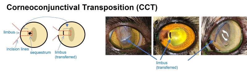
Following the superficial keratectomy, an alternative to a CPG is a corneoconjunctival transposition graft (CCT) surgery, where a thin layer of healthy cornea still attached to conjunctiva is partially dissected from the surface of the eye and then slid into the “crater” left behind where the sequestrum was removed.
The surgeries are performed using an operating microscope, micro-surgical ophthalmic instruments, and with the patient under general anesthesia.
Courtesy of Marnie Ford DVM
A third surgical option after performing a superficial keratectomy is a corneal transplant. This is not performed very often, due to difficulty acquiring donor corneas, and there is a significant risk that the corneal transplant will later develop sequestra.
It is best to not just perform a superficial keratectomy; in the author’s experience, placing a CPG over the cornea best reduces the risk of more sequestra forming later, especially in bug-eyed cats such as Persians.
Potential Outcomes
Owners sometimes opt to not have any surgery performed to remove the sequestrum; they take their chances and hope that Mother Nature will allow the sequestrum to quietly extrude on its own, and the eye will be OK. Sometimes this works out and everyone is happy. But it is a risky roll of the dice, and there can be a very unhappy ending.
It must be emphasized that a “sleeping” well-behaved corneal sequestrum must be thought of as a ticking time bomb. Eventually, the sequestrum WILL usually extrude, and this can happen quietly and slowly and comfortably, or there can be sudden rapid rejection of the sequestrum by the immune system. As the sequestrum extrudes, the melting cornea can rupture and the eye can be lost.
It must be remembered that having a ruptured eye removed still leaves the cat visual in the remaining good eye, because we know that cats with one eye function very well. However, if the second eye develops a sequestrum, it must be aggressively monitored and treated to help reduce the risk of this eye also rupturing and requiring surgical removal.
With surgery, the prognosis for the eye to be free from pain and to have functional vision is good, especially if the sequestrum is removed at an early stage. However, the tendency to form a sequestrum is that individual cat’s response to corneal irritation, and additional episodes may occur in one or both eyes in the future. To help prevent any recurrences, notify your veterinarian immediately if any new eye issues occur.
What happened with Opalina? My clinical findings were as follows:
Diagnoses
- Left eye: Large advanced corneal sequestrum rapidly extruding from the center of the cornea, with secondary acute keratomalacia (corneal melting). I told Maggie that the sequestrum had been forming for months and had been tolerated by the immune system until the past 3-4 days, at which time the immune system suddenly attacked the sequestrum and nearby living cornea, causing the living cornea to melt and the sequestrum to begin sloughing from the cornea. The cornea was going to rupture at any time.
- Right eye: A very early corneal sequestrum. (Once I pointed it out to Maggie with a beam of light, she could barely see the faint golden brown smudge discoloring the center and 3:00 to 6:00 section of the cornea).
- Right eye: Poor quality tear film (the left eye could not be evaluated).
- Right eye: Poor blink (lagophthalmos) that was breed-related (the left eye could not be evaluated as it was squinting severely due to pain).
- Both eyes: Nasal fold trichiasis, with hair contacting the corneal surface. The early corneal sequestrum in the right eye was located where the hair was in contact with the corneal surface.
- Both eyes: Epiphora (tear overflow) due to poor tear drainage (breed-related).
Advertisement
Treatment
I gave Maggie two treatment options for the left eye, but unfortunately medical treatment alone of the left eye was not one of them, because the cornea was too severely affected, Opalina was very painful, and the prognosis was poor.
- Left eye: Emergency surgery to remove the sequestrum and place a tissue graft over the “crater” site where the sequestrum was removed. This had a 60% success rate for Opalina, because the cornea was about ready to rupture and was very sick. But even if the surgery was a success, the area of affected cornea was so large that placing a CPG over that area would cover 80% of the cornea and the graft site would always be cloudy, allowing only 20% vision that is mostly peripheral. I could not perform CCT surgery, because the amount of healthy cornea I needed to harvest and slide into place was larger than what was available to harvest
Note: In a parallel universe, let’s suppose that Opalina had already lost her right eye to a severe corneal sequestrum and now her left eye was in crisis. In this case, surgery to try to save the eye with a superficial keratectomy and CPG is more justified, because even with 80% of the cornea covered with a graft, there is a decent chance that with surgery the eye can be saved, and that 20% of the cornea will be clear enough to provide some vision. A cat can see a lot with just 20% vision!
- Left eye: Emergency surgery to remove the left eye (the eye is removed and the eyelids sutured closed).
- Right eye: No matter what Maggie decided for the left eye, the right eye needed lifetime lubrication with a topical preservative-free lubricant at bedtime, and regular reexamination by the ophthalmologist every 4 months. The best lubricant recommended is OphtHAvet® ointment (Dechra):
- Preservative-free
- Contains hyaluronic acid (HA), which is anti-inflammatory, helps wound healing, increases tear film stability, is soothing, and coats and protects the cornea the longest (as compared with a drop or a gel).
- It is also the only ophthalmic ointment that contains HA
- It is by prescription only, but is available for all veterinarians to prescribe
Maggie’s regular veterinarian did not carry this product, so she obtained it from my clinic and also had a ‘back up’ lubricant handy that she could get online, which is a thicker artificial tear ointment (they are not all created equally). A good one that is thick and coats well is Soothe Nighttime Lubricant Eye Ointment (Bausch & Lomb)
The very best lubricant ointment to apply at bedtime is OphtHAvet® ointment (Dechra), which contains hyaluronic acid (HA) and requires a prescription. The cornea benefits from the long chain high molecular weight glycosaminoglycans in HA molecules, which tangle and stick to themselves and also to the surface of the cornea. HA has many functions, including the following:
- mixes with tears and help reduce evaporation
- stabilizes tear film
- reduces inflammation
- reduces friction (makes it easier to blink)
- absorbs water, to help with corneal hydration
While an ointment is needed for lifetime nighttime lubrication, Maggie was advised that for daytime lubrication, she could apply (as often as needed) a preservative free gel such as Idrop Vet Gel® (I-Med Animal Health) that contains HA and is available online.
What was the end of the story for Opalina? Maggie was not home enough to intensively treat Opalina, and with the guarded prognosis, she was very sad but decided to have the left eye removed.
She was committed to preventing the right eye from having the same fate as the left eye, so I advised her to have a special plastic surgery performed on the inside corner of the right eye at the same time that the left eye was removed, to protect the right eye. This is called medial canthoplasty.
The emergency surgeries were performed the next day, and I combined the medial canthoplasty with partial removal of the nasal fold. The result shortened the inside corner of the eyelid opening, reduced the size of the nasal fold, protected the cornea from hairs rubbing on it, and also improved blinking! But blinking would never be normal. I explained to Maggie that Opalina’s unblinking look of love was mostly because she did not feel the need to blink.
After surgery, Opalina healed well after two weeks in an E collar plus appropriate postoperative medications. She needed lifetime lubrication of the right eye at bedtime, and 1-2x during the day when Maggie was home.
Opalina’s tear staining under her right eye improved after surgery and she blinked better too, which was wonderful. Maggie was forever afterwards vigilant in monitoring the right eye, and took Opalina to see me every 4-6 months for the first year, and then every 6-9 months indefinitely.
Maggie was also advised to contact my office if any squinting or signs of a cold developed, as this could be a recurrent FHV-1 infection with subsequent corneal ulceration and increased the risk of repeat sequestrum formation.
Advertisement
Conclusion
Feline corneal sequestrum is a relatively rare eye condition primarily affecting cats with brachycephalic features such as Persians and Burmese. This condition is characterized by the development of a plaque (sequestrum) that forms within the corneal tissue. The sequestrum is dead corneal tissue that varies in color from light brown to black. The underlying causes of this condition include FHV-1 infection, trauma, corneal exposure, and breeds with a brachycephalic skull shape.
Symptoms are visually apparent and may include corneal discoloration, squinting, and excessive tearing. Diagnosis is most often by a veterinary ophthalmologist, based on clinical appearance. Treatment options vary depending on the severity of the condition and can range from conservative management, such as lubrication and antibiotics, to surgical intervention.
Surgical removal of the sequestrum is usually required to save the eye, prevent further damage to the eye, and alleviate discomfort. Eye removal is sometimes required in severely affected cats. Early veterinary intervention is crucial to manage symptoms effectively and minimize complications. At-risk eyes require daily lifetime lubrication to help prevent formation or progression of corneal sequestra.


By Terri L. McCalla BSc, DVM, MS, Diplomate ACVO
Dr. Terri McCalla Dr. McCalla received a BS in Animal Health Science from the University of Arizona, followed by her DVM degrees from both Oregon and Washington State Universities in 1984. She completed a 3 year residency in Comparative Ophthalmology from the University of Missouri and became board-certified in the American College of Veterinary Ophthalmologists (ACVO) in 1989.