 Pet News

Summary
Ophthalmic examinations are crucial for detecting and managing eye conditions in animals from a young age. Because breed-related longevity challenges age assignment and blurs temporal boundaries. This article focuses on the juvenile patient, this life stage defined as being between the first three months of life and the end of the first quarter of life.
Common eye conditions seen in these young animals include corneal ulcers and uveitis due to trauma, as well as inherited conditions like certain types of eyelid abnormalities and cataracts, and corneal dystrophy. Additionally, polygenic disorders such as third eyelid gland (TEG) prolapse (“cherry eye”), and dry eye syndrome are observed.
The presence of cat claw lacerations, abnormal eyelid hairs causing corneal irritation, and eyelid conformation issues necessitate prompt and appropriate ophthalmologic interventions, ranging from emergency surgery to conservative management with medications or dietary adjustments.
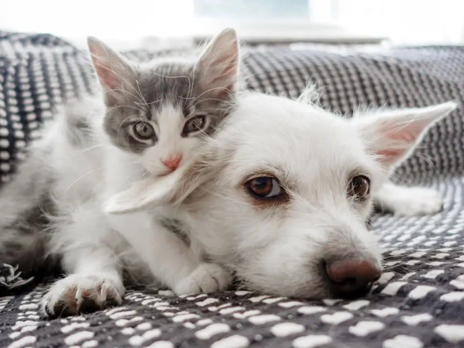
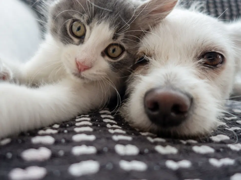
Dr. Emily Rogers peered into the examination room where Max, a lively white mixed-breed puppy, and Allie, a curious little gray and white kitten, were eagerly awaiting their turn with their owner, Rose. As a veterinarian specializing in ophthalmology, Dr. Rogers knew the importance of thorough eye examinations, especially for Rose’s new pets. Rose was very concerned about eye health, having had pets in the past with vision problems, and wanted to be proactive.
Max bounded towards Dr. Rogers as soon as she entered the room, his tail wagging furiously. Allie mewed softly in her pet carrier as Dr. Rogers greeted them and their owner.
“Let’s start with you, Max,” Dr. Rogers said. Her technician scooped up the enthusiastic puppy and placed him on the exam table, gently restraining him. Max squirmed with excitement but soon settled down as Dr. Rogers began her examination.
After the examination, the ophthalmologist explained to Rose that Max’s left eye had a small cataract that was a birth defect and did not significantly interfere with vision, and that these were very unlikely to progress. And that Max’s right eye had a retinal detachment and was not visual. The detachment likely had been present since birth, so Max was born blind in one eye. The right eye was permanently blind. Rose was shocked.
Dr. Rogers explained that Max’s eyes were comfortable, his vision in the left eye was quite functional, and that dogs do great with functional vision in just one eye. She recommended that Max be placed on a special vision supplement to help protect his eyes, in addition to annual eye reexaminations. And to test Max’s vision every day with treats and toys,and to call her office if there was any change in Max’s vision.
Next, it was Allie’s turn. Allie’s eyes were healthy and normal. Dr. Rogers told Rose that Allie did not need to be brought back for ophthalmic reexamination, and to continue regular health maintenance with Rose’s general practitioner veterinarian. But Rose told her that just to be on the safe side, she wanted to bring Allie back for specialist eye examinations every two years, which was fine with Dr. Rogers.
Introduction
Ophthalmic examinations are vital across various life stages, from neonates to seniors, with breed-related longevity differences challenging age assignment and blurring temporal boundaries. This article focuses on juvenile patients, defining this stage as between 3 months of age and first quarter of life. Juvenile patients frequently acquire corneal ulcers or uveitis secondary to trauma. Less frequently, inherited ocular diseases such as eyelid abnormalities, cataracts, and corneal dystrophy, or polygenic conditions such as third eyelid gland (TEG) prolapse and dry eye (keratoconjunctivitis sicca, KCS) can also develop.
Cat Claw Lacerations
Cat claw lacerations frequently occur in young dogs and cats secondary to naivety to claws in combination with an immature menace response. Such injuries often require emergency surgery and should be referred to an ophthalmologist for urgent care. It is important to remember that young animals have neither the wherewithal to understand that other animals can hurt them, nor the reflexes to get out of the way! Keeping a watchful eye on young patients, especially when around older animals will help to keep them safe.
Advertisement
Abnormal Eyelid Hairs

Abnormal eyelid hairs are more common in dogs than in cats and range in severity and between breeds. A focal superficial ulcer identified in a juvenile dog suggests the presence of an ectopic cilium as the cause of the ulcer. Ectopic cilia are extremely rare in cats. Ectopic cilia form when an abnormal hair emerges through the palpebral conjunctiva to directly poke the surface of the cornea. Removal of the offending hair using high magnification but requires a very short general anesthetic. The hair follicle must also be removed to prevent regrowth of the hair. Other commonly found abnormal hairs that can contact the cornea are distichia. Distichia are hairs that protrude through the meibomian gland openings that border the eyelid margin.
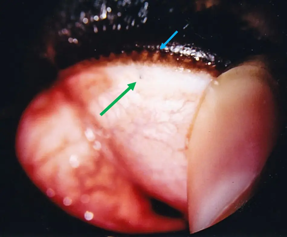
Distichia vary in length, stiffness, and number but rarely do they directly cause corneal ulcers. Ulcers that develop secondary to distichia are typically caused indirectly by irritation and self injury by rubbing. Removal of distichia is recommended in cases of irritation and ulceration. Misdirected eyelashes, or eyelid or nasal fold hairs that contact the cornea, are called trichiasis . Trichiasis are commonly seen in brachycephalic breeds with macropalpebral fissure syndrome whereby the hairs from the nasal folds contact the cornea. Chronic irritation by these hairs may cause pigment to become deposited onto the cornea. If progressive, the pigment can cover the whole cornea to impair vision or even blindness. Early evaluation, management, and surgical correction of eyelid conformation by an ophthalmologist are recommended when the amount of corneal pigment is noted to be increasing.
Third Eyelid Gland Prolapse “Cherry Eye”

Normally hidden, the TEG is anchored to the orbital wall by ligaments. The gland is most commonly prolapsed in young dogs and rarely in cats and is aesthetically unpleasing. When prolapsed, this gland often becomes enlarged, which can not only interfere with blink function, but can become easily traumatized and predispose the patient to developing dry eye later in life. Prolapsed TEG may be mildly uncomfortable when it first occurs. Surgical replacement of the gland is recommended in an effort to preserve tear function of the gland. For more information about third eyelid gland prolapse or “cherry eye” please visit the article entitled “Third Eyelid Gland Prolapse in Dogs”.
Advertisement
Keratoconjunctivitis Sicca, KCS
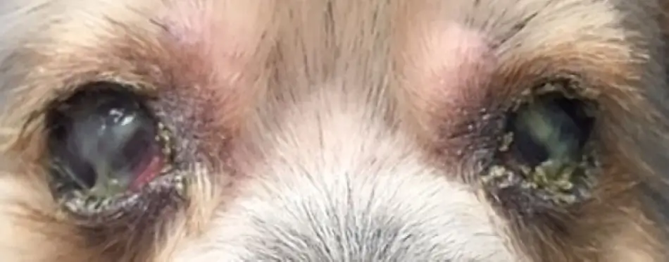
KCS can be described in two ways: poor tear volume (i.e., quantitative tear film deficiency) as measured by the Schirmer Tear Test (STT), or reduced tear film quality (qualitative tear film deficiency) as diagnosed by an ophthalmologist using the Rose Bengal dye test and measurement of tear film breakup time (TFBUT). The STT should be measured when persistent mucoid discharge is noted, and only when nothing has been applied to the eyes on the morning of the examination to avoid confounding the results with fluid added. Some patients can have KCS due to qualitative tear film deficiency and yet have normal STT values. KCS can cause secondary bacterial conjunctivitis, discomfort, ulcers, and pigmentation.
The causes of KCS are numerous but most commonly occur secondary to immune-mediated destruction of the tear glands or to congenital absence of the lacrimal gland(s). Dry eye is extremely uncommon in cats. Treatment of KCS is most commonly achieved with the application of a topical tear stimulant, sometimes supported with topical antibiotics and lubricants. It is important for the health of the eyelids and corneas to keep the eyes and eyelids free of accumulating ocular discharge. For more information, please see the Related Articles to this article.
In severe cases that do not respond to medical therapy, a parotid duct transposition surgery may be performed with mixed success. For more information about dry eye, please visit the article entitled Dry Eye in Dogs.
Eyelid Abnormalities
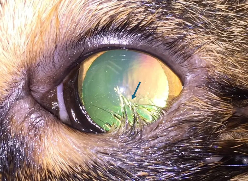
Eyelid abnormalities such as entropion, ectropion, and lagophthalmos (incomplete blink) are polygenic conditions which are congenital or develop as the head grows and/or the facial skin becomes heavier. Entropion may be spastic due to squinting and globe retraction or, as with ectropion, can develop secondary to eyelid injury and secondary scarring (cicatricial). Entropion is exacerbated with deep set eyes, corneal pain, or macropalpebral fissure syndrome.

Temporary tacking sutures that evert the eyelid may be placed, but permanent correction is ideally not performed until the patient has reached adult head conformation. To reduce irritation associated with eyelid hair contact with the cornea, application of a corneal lubricant several times daily is recommended until tacking sutures can be placed. Ectropion and lagophthalmia may be normal features in some dog breeds and may not require surgical correction. For more information about entropion, please visit the article entitled Entropion – An Overview.
Cataracts
Cataracts may be unilateral or bilateral and can vary in time of onset and size between the two eyes. Most cataracts seen in the juvenile patient are inherited, and all cataracts should be evaluated by a veterinary ophthalmologist to determine if or when surgical removal is indicated. If surgery is not elected, many patients require therapy for inflammation in the eye that is caused by the cataract (lens-induced uveitis). You can read more in our article on Cataracts.
Corneal Dystrophy

Corneal dystrophy is inherited, often slowly progressive, typically present on both eyes centrally, and not associated with prior corneal disease. The deposits are typically calcium or fat. Your veterinarian can perform some blood tests to determine if high cholesterol, triglycerides, or calcium, or low thyroid values are present. Any changes noted can usually be addressed with dietary changes or daily medication. This condition is not reported to be irritating; however, irritation is more often associated with calcium deposition (spiky crystals versus the soft deposits of fat), and a medication can be prescribed to help blunt these sharp calcium points. Until a prescription can be provided to remove/blunt these deposits, application of a corneal lubricating gel is recommended.
Advertisement
Conclusion
In juvenile animals, timely ophthalmic evaluation and intervention are paramount for preventing long-term visual impairment due to a variety of congenital, inherited, and trauma-induced eye conditions. Proactive measures, including surgery for structural abnormalities like TEG prolapse and entropion, alongside medical management for conditions such as KCS and cataracts, are essential. This comprehensive approach not only alleviates immediate discomfort but also addresses potential complications, thereby enhancing the quality of life and preserving the vision of these young patients. Early detection and treatment are critical, especially given the susceptibility of juvenile animals to rapid progression of ocular diseases and their limited ability to avoid harm.
After completing their examinations, Dr. Rogers sat down with Rose, as Rose was upset about Max’s right eye being blind. Dr. Rogers reminded her that Max did not know any difference- to him, this was normal. Dogs have amazing hearing and sense of smell, and compensate very well. It was fairly common for ophthalmologists to diagnose blindness in one eye in a cat or dog, and the owner to not have a clue that vision loss was present.
Rose was so grateful that she had brought in her new pets to have them checked out. She told Dr. Rogers that she would be back next year with Max, and would start him on a vision supplement as soon as possible.
Dr. Rogers couldn’t help but smile as the trio left her clinic. Another day, another opportunity to make a difference in the lives of her patients, one wagging tail and purring kitten at a time.
By Marnie Ford PhD, DVM, Diplomate ACVO
Dr. Ford graduated from the Ontario Veterinary College in 2000 after completing a Bachelors in Zoology at the University of British Columbia and a PhD in Physiology at Monash University in Australia. Following a rotating small animal internship at the University of Minnesota in 2001, Dr. Ford achieved Diplomate status from the American College of Veterinary Ophthalmologists in 2006 after completing a 3-year residency in Comparative Ophthalmology at the University of Missouri. After co-founding a private referral hospital, West Coast Veterinary Eye Specialists, in New Westminster (2004-2013) she opened Pacific Animal Eye Specialty services, providing mobile medical and surgical ophthalmologic care to patients in the Lower Mainland, Vancouver Island, and the Interior of BC (2013-2019). In 2019, she returned to Australia where she worked full-time in both medical and surgical ophthalmology as well as resident training at Animal Eye Care in Melbourne Australia. In 2023, Dr. Ford returned to Vancouver to provide locum services to ophthalmologists across Canada.