 Pet News


Summary
The indolent ulcer, known by various other names, was first documented in veterinary medicine in 1954. Since then, cases have been reported in cats, dogs, and horses. This condition is also common in humans. These chronic, superficial corneal ulcers resist normal wound-healing processes and predominantly affect middle-aged to older animals, with rare occurrences in younger individuals.
Changes that occur include shallow ulcers with loose epithelial edges and a faint ring around them following application of fluorescein dye. Treatment strategies can be challenging and are best approached with the guidance of a veterinary ophthalmologist. The aim of therapy is to remove non-adherent epithelium and promote healthy epithelial regeneration.
Steve loved Boxers and had a sweet eight year old female named Bella. One day Bella’s left eye started watering and she favored it slightly. The next morning, she kept her left eye closed and pawed at it. Steve was able to get an appointment later that day with his veterinarian, Dr. Drake, who diagnosed a superficial corneal ulcer.
Bella was placed on a topical antibiotic eye drop and an oral pain medication, plus an Elizabethan collar (E collar—AKA the “cone of shame”). Steve hated the cone and felt terrible putting it on Bella, so after one day he took it off. But the eye was not healed at recheck 5 days later, and the ulcer was even larger (but not deeper). On went the E collar, 24/7. But over the next several weeks, the cornea did not heal. Dr. Drake had even performed a “Q Tip debridement”, in which he numbed the cornea with eye drops and removed non-healing loose tissue from the surface of the cornea with a sterile cotton-tipped applicator, in hopes that the tissue would regrow correctly and heal the ulcer.
Finally, after one month of Bella not getting any better, Dr. Drake referred her to a veterinary ophthalmologist, Dr. Mason. The earliest available appointment was two weeks later.
Steve was very frustrated and upset that his Bella had been in pain and in a cone for so long, in spite of the time and money spent on three veterinary visits. He was worried that Bella might lose her eye and anxiously waited the next two weeks until Bella could be seen by the specialist.
Introduction
Indolent ulcers in dogs, also known as superficial non-healing corneal ulcers, spontaneous chronic corneal epithelial defects (SCCEDs), chronic corneal erosions, refractory ulcers, or Boxer ulcers, manifest as chronic, superficial wounds that defy typical healing mechanisms. These ulcers, identified in dogs, cats, and horses, primarily affect middle-aged to older animals, persisting for weeks to months if improperly managed.
Notably, affected animals often lack concurrent ocular diseases like keratoconjunctivitis sicca (KCS, dry eye), ectopic cilia, or eyelid entropion. The condition is rare in young animals, with older Boxers and middle-aged French Bulldogs exhibiting a disproportionate prevalence. These ulcers resist conventional treatment, highlighting the necessity for tailored therapeutic interventions to promote effective healing and alleviate discomfort.
Advertisement
Cause
Arising from minor eye injuries that typically heal promptly in younger animals, indolent ulcers can be notably sluggish in their healing process, often resulting in discomfort and additional changes like corneal blood vessel infiltration (vascularization) and edema. These ulcers are shallow with loose epithelial edges and a faint ring around them that becomes apparent after applying fluorescein dye.
When examined microscopically, the outermost layer of the cornea (the epithelium) is noted to be made up of abnormally shaped cells which lack normal adhesion complexes that keep these cells attached to the inner layer of the cornea called the stroma.
In addition to a lack of adhesion complexes, a thin membrane called hyalin forms on the surface of the stroma which further obstructs healing. For these reasons, treatment approaches are aimed at removing any abnormal epithelium and hyalin and promoting the formation of healthy epithelium and its adherence to the stromal layer of the cornea.
Diagnosis

Clinical hallmarks of indolent ulcers include superficial, non-infected corneal ulceration with loose epithelial edges and mild corneal edema, often accompanied by variable ocular pain and delayed corneal vascularization. Ocular pain, which tends to diminish with time, is seen as squinting (blepharospasm), tearing (epiphora), and rubbing at the eye or face.
Application of fluorescein dye to the cornea by your veterinarian reveals a halo of loose epithelial tissue seen as less-intense staining, surrounding the more intensely stained corneal ulcer. Indolent ulcers can occur anywhere on the cornea, have mild corneal edema localized to the lesion site, and often have corneal blood vessels, especially when the ulcer is present on the periphery of the cornea instead of in the central zone.
The diagnosis of an indolent ulcer should be considered in any middle-aged to older dogs presenting with non-healing ulcers lasting at least 1-2 weeks. A thorough eye examination is still essential to rule out underlying causes of delayed wound healing, including mechanical trauma, foreign bodies, infections, tear film abnormalities, or structural abnormalities, particularly in younger dogs.
Advertisement
Etiology

The cornea comprises four distinct layers, each with unique cellular makeup and organization. The top two layers (the epithelial layer and superficial stromal layer) are affected by indolent ulceration. Normal small, superficial ulcers typically heal within 24 to 72 hours through normal cell division, migration, and adhesion.
For indolent ulcers, the normally rapid healing process is slowed down or even restricted by the failure of the epithelium to produce normal basement membrane and adhesion complexes, resulting in disruption in epithelial adherence to the stroma.
Unlike in humans, indolent ulcers in dogs are not associated with specific congenital eye disorders. Treatments aim to address these tissue-level changes to help ulcer healing.
Treatment
Many medical and surgical therapeutic options have been described for the treatment of indolent ulcers. The therapy chosen is based on the observed changes, the experience of the veterinarian, owner compliance, and financial aspects. It is important to understand that multiple treatments might be necessary to heal these ulcers, and that recurrence in the same or opposite eye is possible.
Various treatment options exist for indolent ulcers, and they are typically each followed with prophylactic antibiotics recommended to prevent secondary infections of the cornea, a medication to dilate the pupil for comfort, and an Elizabethan collar to prevent self-trauma. Treatments and their effectiveness vary, and so treatment choices depend on clinician preference and experience.
Medical therapy is aimed at reducing pain and infection and includes antibiotics, hypertonic saline, lubricants, and pain medications. Less commonly used medical treatments use substances like epidermal growth factor and aprotinin.
Surgical approaches involve removing loose epithelium and abnormal basement membrane and promoting new epithelial growth with strong adhesion complexes. Surgical interventions range from debridement, superficial keratotomy, superficial keratectomy, or less conventional surgical approaches that include application of tissue adhesive, placement of a third eyelid flap, thermal cautery, or placement of a conjunctival graft.
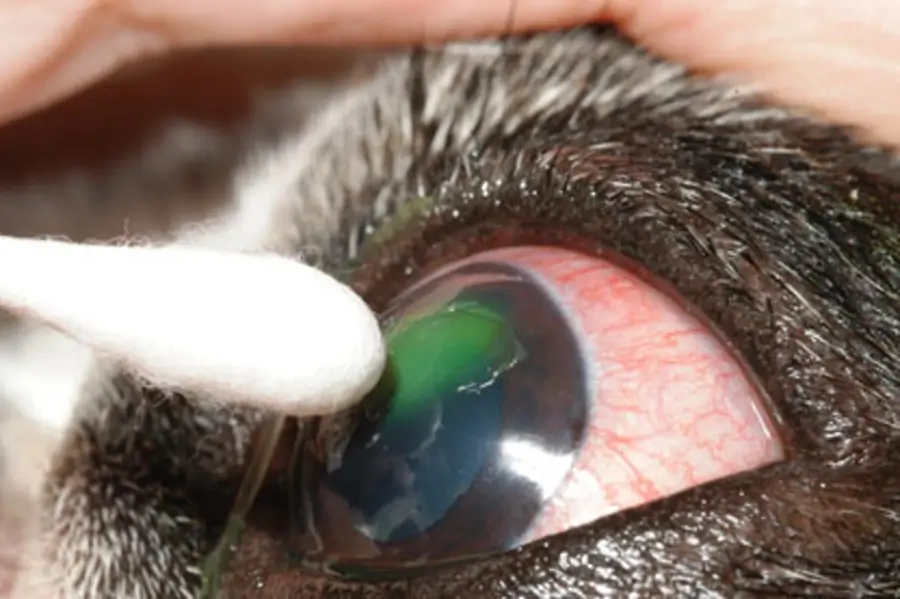
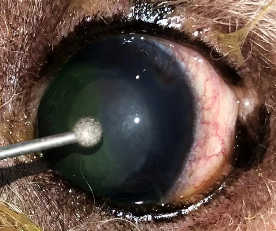
Corneal debridement is typically the first surgical approach to an indolent ulcer. After the cornea has been numbed using topical anesthetic drops, cotton-tipped applicators or a diamond burr device is used to wipe off or gently scrape (debride) loose edges of epithelial tissue from the corneal surface.
This procedure not only cleans the cornea of loose epithelial tissue incapable of sticking to the underlying stroma, but also helps to remove some of the hyalinized membrane, if minimally present, by creating micro-erosions of the basement membrane, promoting the assembly of epithelial cell adhesion complexes, and enhancing the adhesion of new epithelial
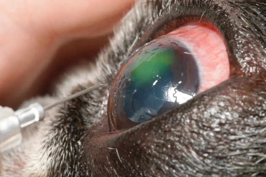
A keratotomy is performed by creating very superficial, linear scratches (grid keratotomy, striate keratotomy), or pinpoint cuts (multiple punctate keratotomy) in the surface of the stroma.
By doing so, the healthier superficial stromal tissue at each keratotomy site is exposed and can more easily allow the newly growing epithelial tissue to stick.
A keratotomy should never be performed on a cat or horse as this can result in other disease conditions that include corneal sequestrum formation (cats) or fungal infection (horses).
A keratectomy is the removal of normal and abnormal tissue. Unlike corneal debridement which removes epithelial tissue that was not actually adhered to the underlying stroma, a keratectomy involves removal of some of the healthy tissue immediately underlying the unhealthy epithelium and superficial stroma.
During this procedure, typically only the top ½-1/3 of the cornea is removed, leaving a fresh and healthy tissue bed for new epithelium to grow over and heal. Superficial keratotomy or keratectomy is believed to facilitate healing by removing abnormal stromal membranes and promoting epithelial adhesion to the corneal stroma, possibly through strengthening the adhesion of the epithelium to corneal collagen fibers. However, the precise mechanisms underlying these processes is not fully known.
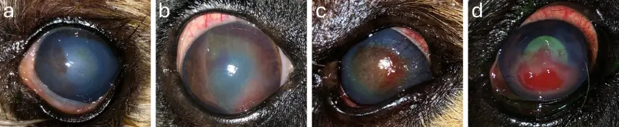
Following any surgical procedure, adjunct therapies can be provided to increase comfort and provide corneal protection against irritation by the eyelids. These therapies include application of a soft contact lens or placement of a temporary eyelid suture (tarsorrhaphy) to keep the eyelids partially closed over the sensitive corneal during the healing process.
It is common for a cornea to heal with an infiltration of blood vessels. These vessels may be sparse or robust and extending into all off or only part of the original ulcer site. While these blood vessels may appear alarming, they are normal and help with corneal healing. The blood vessels will regress slowly over time.
A less commonly used surgical option includes placement of a conjunctival graft. Conjunctival grafts are only indicated in cases of deep ulcers or in cases in which other therapies for indolent ulcers are not successful.
Recurrence
Indolent ulcers may recur due to underlying abnormalities. Following corneal ulceration that is not indolent, we know that the cornea can take up to a year for the healed epithelial tissue to be adhered to pre-ulceration strength.
From this, it has been suggested that corneas of patients with indolent ulcers are abnormal before the indolent ulcer occurs and that the corneal tissue remains compromised for a longer period following apparent resolution. For these reasons, these corneas remain prone to recurrences.
Advertisement
Conclusion
Indolent ulcers pose a significant challenge in veterinary ophthalmology, requiring a comprehensive understanding of their etiology, clinical presentation, and treatment options. Successful management relies on accurate diagnosis, appropriate medical and surgical interventions, and an understanding of the potential for recurrence and the importance of follow-up care.
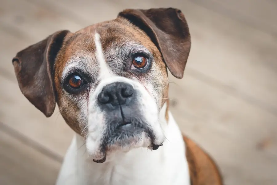
Dr. Mason told Steve that Bella had an indolent ulcer, which was very common in older Boxers. These were also common in any older dog, and occurred when there was a very superficial break (erosion) in the surface layer (epithelium) of the cornea that failed to heal within two to three days because the epithelium was defective.
He explained that a very successful minor surgery could be performed under mild sedation and a topical numbing drop to get the cornea to heal. The surgery involved removal (debridement) of the epithelium, then “roughening up” the underlying cornea so that new epithelium could regrow and properly stick down. This could be accomplished by using a tiny needle, or by burring the surface—somewhat like sanding wood before painting it, in order for the paint to stick.
Dr. Mason used a device called a diamond burr. Then a clear therapeutic soft contact lens was placed as a bandage to protect the healing cornea and improve comfort. Bella would need to continue to wear an E collar until recheck in two weeks, but the one she was wearing was way too short and was the wrong style, so she’d need a new one that was custom-fit because she must not be able to rub her eye even once, as this would peel off the new epithelium before it had a chance to stick and anchor itself down by forming microscopic attachments. And that nearly all corneas are healed at recheck two weeks after surgery.
Dr. Mason explained that because all of the epithelium was defective, all of the corneal epithelium needed to be removed. This would greatly reduce the risk that Bella would later get another nonhealing indolent ulcer in this eye, in an untreated area. And that all topical medications needed to be preservative-free, because preservatives damaged the fragile newly growing epithelium– especially benzalkonium chloride.
So Bella had the procedure that same day and was discharged with her new custom cone and new topical medications, plus continuing the pain medication. Her cornea was healed two weeks later, and Steve was overjoyed. No more cone and no more medications!
But two years later when Bella was ten, her right eye started squinting and tearing. Steve knew the drill and went into action. At the recommendation of Dr. Mason’s staff, Steve had kept Bella’s custom E collar, so he popped it on her head and called Dr Mason’s office.
Dr. Mason’s staff was not surprised at the news, and was able to schedule Bella two days later to be examined, plus extra time for a surgical debridement and keratotomy procedure. Dr. Mason confirmed that an erosion was present and that the epithelium did not look very healthy, so Bella underwent her second—and final— corneal surgery. The cornea was healed two weeks later!
Steve tossed the E collar in the plastic recycling bin, and the man and his dog lived happily ever after.
By Marnie Ford PhD, DVM, Diplomate ACVO
Dr. Ford graduated from the Ontario Veterinary College in 2000 after completing a Bachelors in Zoology at the University of British Columbia and a PhD in Physiology at Monash University in Australia. Following a rotating small animal internship at the University of Minnesota in 2001, Dr. Ford achieved Diplomate status from the American College of Veterinary Ophthalmologists in 2006 after completing a 3-year residency in Comparative Ophthalmology at the University of Missouri. After co-founding a private referral hospital, West Coast Veterinary Eye Specialists, in New Westminster (2004-2013) she opened Pacific Animal Eye Specialty services, providing mobile medical and surgical ophthalmologic care to patients in the Lower Mainland, Vancouver Island, and the Interior of BC (2013-2019). In 2019, she returned to Australia where she worked full-time in both medical and surgical ophthalmology as well as resident training at Animal Eye Care in Melbourne Australia. In 2023, Dr. Ford returned to Vancouver to provide locum services to ophthalmologists across Canada.