 Pet News

Summary
Progressive Retinal Atrophy is the most common retinal disease in dogs. It is an umbrella term for a group of genetic diseases that affect many breeds of dogs, causing gradual retinal degeneration, secondary toxic cataracts, and eventual blindness in both eyes. It is recessively inherited, with some breed-specific exceptions. The disease initially causes night-blindness and dilated “glowing” pupils, progressively leading to complete vision loss. Although there is no treatment for PRA, there is hope for slowing down vision loss. Advancements in understanding retinal damage and oxidative stress have led to promising supportive care for retina and lens health, which aim to slow the degeneration process to preserve vision for a longer period. Early detection is crucial, and affected dogs should not be bred. After the diagnosis, regular examinations by a veterinary ophthalmologist are needed for optimal management and care.
Robbie was a seven year old Yorkshire Terrier with pet parents Jill and Dave, who adored him. Robbie loved to play with his red ball and sit at the big picture window to watch for birds and cats in the yard, barking madly whenever he saw an invader. Dave took him everywhere in his truck. Everyone knew Robbie and he got treats at the bank drive-thru, neatly catching the treat every time one was tossed. But lately, he was getting clumsy and missing some of the treats.
And then one overcast evening, Robbie kept missing the ball when Jill and Dave threw it. Dave and Jill looked closely at Robbie and noticed that his eyes looked glassy. They tossed him a treat. He missed it but heard it land and readily found it.
Shaken, Dave and Jill had Robbie examined by their veterinarian, who explained that Robbie appeared to be healthy other than his vision. Retinal disease was suspected, and Robbie was referred to a veterinary ophthalmologist.
An appointment was made with a veterinary ophthalmologist but the earliest that Robbie could be seen was in 3 weeks. In the meantime, their veterinarian suggested that Dave and Jill keep a log of whenever Robbie made any visual mistakes, and under what circumstances. And to also watch for any other abnormalities, such as seizures or poor balance (ataxia) or anything else.

Introduction
Progressive retinal atrophy (PRA) is an umbrella term for a large group of inherited retinal diseases in dogs, in which the entire retina thins and degenerates over time, causing gradual loss of night vision followed by loss of day vision, and eventual blindness. In fact, if a dog was destined to lose vision and the author could pick how the dog would become blind, PRA would be the top choice. PRA is not painful, the slow loss of vision allows the dog to gradually adapt to their reduced vision, and there are strategies to help slow down vision loss and also to help the dog cope.
The retina is the light-sensitive “film in the camera” layer at the back of the eye, and is responsible for converting light into electrical signals, which the brain interprets as vision. The retina contains that are intricate cells that detect light and transform it into electrical energy, which in turn is sent by the optic nerve to the brain, where vision takes place.
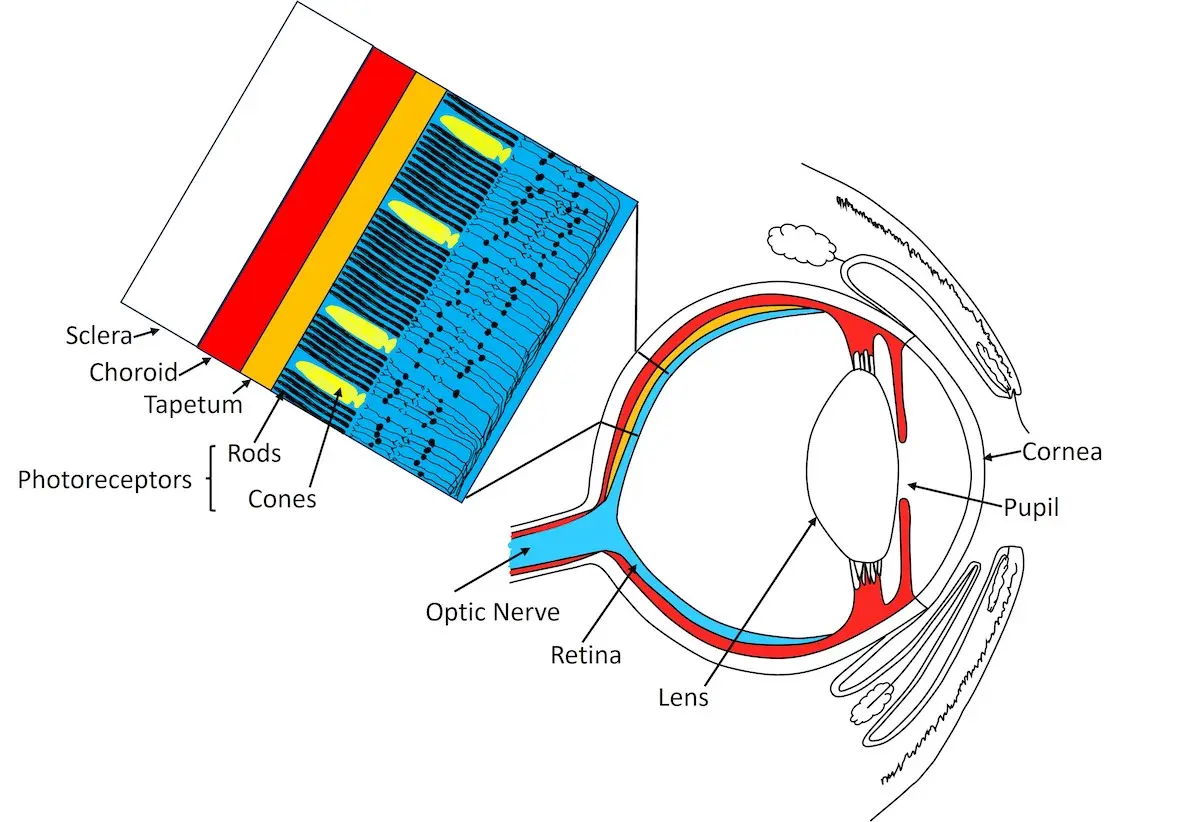
The vast majority of photoreceptors (95%) are rods, which help with vision in dim light and with motion detection. Cones help with vision in bright light and with color vision. In the most common types of PRA, rods are targeted to die, followed by secondary slower cone death. Thus, PRA initially kills 95% of the photoreceptors! This occurs in both eyes simultaneously and is nonpainful. Progressive retinal atrophy occurs in most breeds of dogs and also occurs in mixed breeds.
Some types of PRA are comparable to retinitis pigmentosa in humans; studying PRA in dogs has also contributed to a better understanding of similar conditions in people. While there are dozens of inherited retinal degenerative diseases in dogs, discussing each of these in detail is beyond the scope of this article.
Advertisement
Causes
The retina is derived from the same type of embryonic cells as the brain, and these two tissues have the highest oxygen demand in the body. Rod and cone cells require a rich blood supply, but too much oxygen is toxic and causes cell damage due to an imbalance of both antioxidants and the unstable byproducts of oxygen metabolism (free radicals). It has been theorized that the death of rods by PRA results in too much “left-over” oxygen, which causes the slower death of cones.
PRA can be classified three ways: 1) the type of gene mutation; 2) the mode of inheritance; and 3) the age of onset.
Progressive retinal atrophy is primarily caused by recessively inherited genetic mutations, resulting in photoreceptor degeneration and death. Most mutations occur in rods rather than cones. Different breeds have specific genetic markers associated with PRA, making genetic testing an essential tool for breeders. The most common form of PRA is late-onset. It occurs in over 50 breeds and is a mutation in the PRCD (Progressive Rod-Cone Degeneration) gene, resulting in a mutated protein that is important in the health of rods. Thus, the death of photoreceptors in PCRD is rod-led, with secondary slower death of cones.
Dogs with PRCD are born with normal rods and cones, but the cells die in adulthood, gradually leading to blindness. In most types of PRA, because rods die first, night vision is first affected. Rarely, dogs can have a form of PRA in which cones die first. This is called day blindness (achromatopsia), and occurs in Siberian Huskies, German Shepherds, Malamutes, and Labrador Retrievers.
A less common form of retinal disease occurs in puppies who are born with rods and/or cones that never develop properly, which leads to blindness much more quickly. Eventually, all genetic variations of PRA progress to blindness.
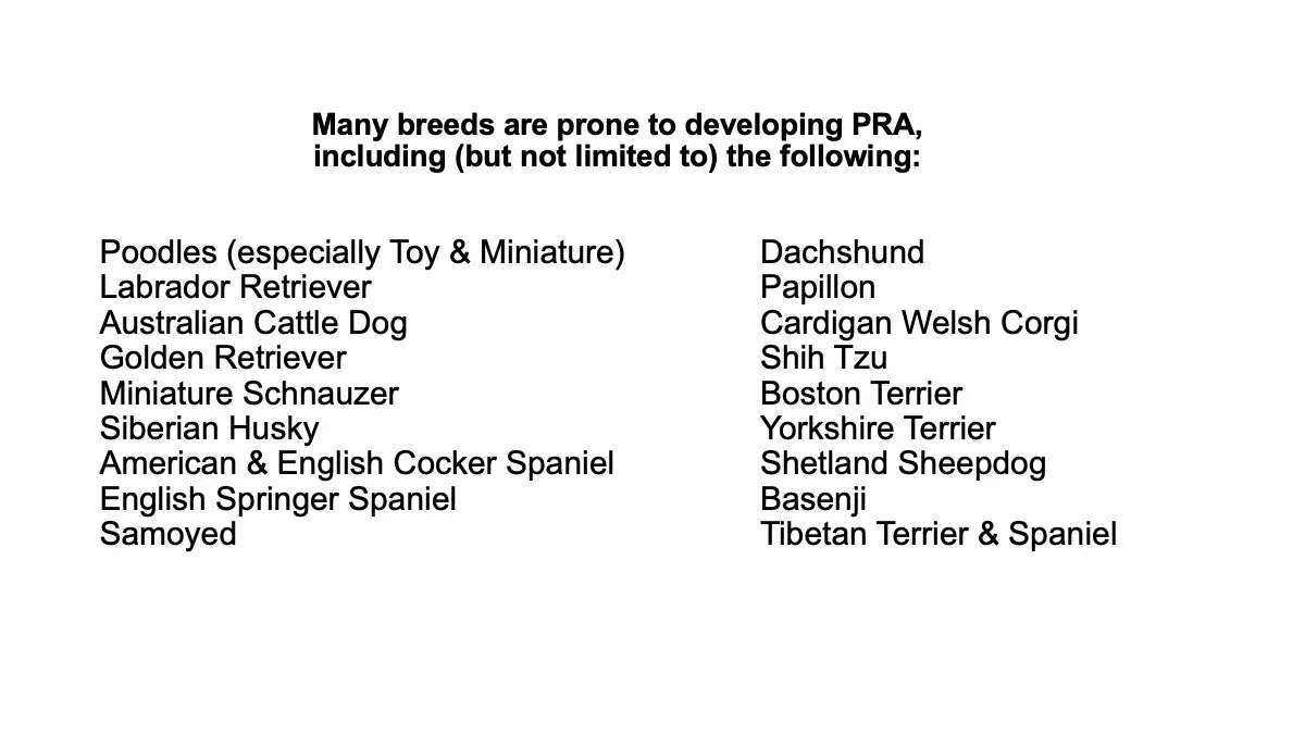
PRA can be classified in over 20 forms based on genetic mutations. PRA is recessively inherited in all breeds studied except it is dominantly inherited in Old English Mastiffs and Bullmastiffs, and is sex-linked and found primarily in male dogs in Siberian Husky, Samoyed, and Weimaraner breeds.
A special note about Mastiffs and Bullmastiffs affected with PRA: their rods are killed by light levels that are safe for normal dogs, so the rods are very sensitive to light and affected dogs with greater exposure to light lose their vision faster. When examining the eyes of a Mastiff or Bullmastiff in which PRA is suspected, minimum light levels should be used. Pet owners should take precautions when shining lights into their dog’s eyes, taking flash photos of their dog, and if the dog lives where light levels are naturally high. These dogs would also benefit from wearing protective goggles.
When classified by the age of onset, PRA can be divided into two types: early-onset and late-onset PRA. Early-onset PRA includes a form of inherited retinal dysplasia that appears in puppies as young as a few weeks old. All rods and cones in the retina never fully develop and then degenerate. This condition progresses rapidly, often leading to blindness within the first year of life, Breeds commonly affected by this form of PRA include the Cardigan Welsh Corgi, Collie, and Irish Setter. Another variation of early-onset PRA is inherited retinal dystrophy (degeneration), where the retina develops normally at first but begins to degenerate early in the dog’s life. This type of PRA is seen in breeds such as the Papillon and American Pit Bull Terrier.
Late-onset PRA is more common and includes forms involving the PRCD gene. This type of PRA is characterized by inherited retinal dystrophies that typically manifest in adult dogs between the ages of six and nine years, though it can occur as early as three years of age. The progression of late-onset PRA is slower compared to the early-onset type.
Incidence
The true incidence of PRA in various breeds is unknown, as there is no centralized health database. The incidence of PRA among breeds often varies geographically, where some breeds may be more popular than others. While professional and ethical breeders maintain responsible breeding programs and test their breeding dogs for various health conditions, “hobby breeders” do not typically test their breeding animals for genetic health conditions, allowing the amplification and spread of inherited conditions in the dogs that they sell. For example, if Australian Cattle Dogs are more popular locally, then more dogs will be bred by hobby breeders and any bad genes amplified.
Clinical Signs
The clinical signs of PRA are generally progressive. The age of onset and rate of progression can vary, even between dogs with the same genetic type of PRA. Because PRA most commonly makes rods die first, and rods are responsible for vision in dim light (“night vision”), the first clinical signs that the owner often notices are night-blindness (poor vision in dim light) and that the dog is reluctant to go outside at night and to enter unfamiliar dimly-lit areas. Both pupils are dilated and owners often notice a “glow” or increased “eye shine” from the eyes.
Clinical signs in dogs with PRA vary from the dog first becoming night blind in the early stage of PRA, to the entire visual field in all light levels becoming affected in advanced PRA.
In the early stages of PRA, clinical signs often include night blindness (nyctalopia), where dogs experience difficulty seeing in low light or darkness. This condition is typically observed when the dog hesitates to move in dim lighting, avoids going outside at night, or misses tossed treats or toys in low-light conditions. Another sign is dilated pupils where both pupils appear larger than normal, even in bright light.
Although the pupils may still constrict in response to bright light, they usually do so more slowly than normal. Additionally, there is an increase in eye shine, a phenomenon where the tapetum, a reflective layer beneath the retina, becomes more hyperreflective due to retinal degeneration.
This degeneration thins the retina, creating the appearance of brightly glowing eyes in dogs affected by PRA. This effect is similar to a layer of aluminum foil (the tapetum) being covered by a layer of tissue paper (the retina).
If the tissue paper degenerates and thins to the point of near transparency, the foil underneath becomes super-shiny and “hyperreflects” through the tissue paper.

Dr. David Ramsey Photo
Fig 5. Video of a dog with PRA. Both pupils are widely dilated. Note the eye shine as the dog turns its head.
In the more advanced stages of PRA, clinical signs become more pronounced and include progressive vision loss, eventually leading to blindness in all lighting conditions. Dogs with PRA may exhibit a “searching” wide-eyed gaze as they rely more heavily on their hearing and sense of smell to navigate their environment. Another development in up to 27% of dogs with more advanced PRA is the occurrence of secondary toxic cataracts in both eyes.
These cataracts form when toxins released from dying rod and cone cells diffuse into the vitreous and come into contact with the back of the lens, causing oxidative damage. The resulting cataracts typically worsen over time, further impairing the dog’s vision, and can prevent early diagnosis of PRA by interfering with pet parents noticing increased “eye shine” (tapetal hyperreflectivity). In some cases, dogs are not diagnosed with PRA until after they have developed advanced secondary cataracts, a diagnosis that requires specialized testing by a veterinary ophthalmologist.
In the natural course of the disease, most dogs will develop blindness within one year of diagnosis. This often occurs because many affected dogs are not diagnosed with PRA until the disease is more advanced, as dogs are quite adept at “hiding” their reduced vision by using their keen senses of hearing and smell to navigate their surroundings. Because dogs with PRA compensate so well for their gradual vision loss, it is very easy for pet parents to not notice any clinical signs, including vision loss, until the PRA is advanced.
Advertisement
Diagnosis
Diagnosing PRA requires referral to a veterinary ophthalmologist for ophthalmic examination and clinical evaluation. Veterinary ophthalmologists have the expertise and the specialized diagnostic equipment to best diagnose retinal disease in animals.
Key diagnostic steps for diagnosing PRA in dogs begin with a thorough review of the dog’s medical history and an observation of clinical signs that may suggest PRA, such as changes in pupillary light reflexes and the dog’s ability to track moving objects like treats or cotton balls. The next step involves an ophthalmic examination, where the veterinarian examines the fundus, which includes the optic nerve, retina, retinal blood vessels, and tapetum, using ophthalmoscopy. This examination helps identify signs of retinal degeneration and any secondary degeneration of the optic nerve. Additionally, slit lamp biomicroscopy is used to examine the anterior segment of the eye to detect secondary toxic cataracts that may develop as a result of PRA.
To further assess the function of the rods and cones in both bright and dim light, electroretinography (ERG testing) is sometimes employed, but is usually not required if the fundus is able to be visualized and has the typical appearance associated with PRA. This tool is particularly valuable in diagnosing PRA when the fundus cannot be directly examined due to advanced secondary cataracts. Genetic testing is another crucial component, as DNA tests can identify specific genetic mutations associated with some forms of PRA. These tests are breed-specific and can help pinpoint carriers and affected dogs. However, it is important to note that not all forms of PRA have identifiable genetic markers, and a dog testing negative for PRA on a genetic test does not entirely rule out the disease. The two most popular DNA test kits are Embark® and Wisdom Panel™.
These DNA test kits are particularly useful for identifying young affected purebred dogs before they exhibit clinical signs of PRA, allowing them to be removed from breeding programs to prevent the gene from being passed on to future generations. Early diagnosis through DNA testing also enables affected dogs to receive specific daily oral antioxidants early in the disease course, which supports retinal health by reducing oxidative damage, potentially slowing the death of rod and cone cells, and reducing the risk of secondary toxic cataracts. However, if PRA is diagnosed by an ophthalmologist through clinical examination, genetic testing is not typically performed unless a breeder desires testing.
What to do if you suspect your dog has PRA
It is important to get an accurate diagnosis. Have your dog examined by a board-certified veterinary ophthalmologist to determine if PRA is present. For a list of board-certified veterinary ophthalmologists, please visit https: www.acvo.org. Dogs with PRA should not be bred, and the breeder that you received your dog from should be notified of this diagnosis so the breeder can alter their breeding program.
Advertisement
After the Diagnosis
Don’t give up! PRA is no longer a hopeless disease. Please read Part 2 and Part 3 of this three-part series about PRA, to learn about treatment, management, and potential outcomes of PRA in dogs.
While it is normal to grieve about your pet’s vision loss, you must not put your sad feelings in your dog’s head—they aren’t there! Your dog’s job description has not changed, and they are happy as long as their routine is stable. From your dog’s point of view, life continues to be great– they just need to use their other keen senses a bit more. Keep household furniture in its place, and consider purchasing the book “Blind Dogs and Seeing Eye Humans: Unconditionally loving and training a blind dog”.
It is also important to know that after your dog is diagnosed with PRA, if their vision is rapidly declining you must contact your veterinary ophthalmologist’s office. This new vision loss may be due to rapid secondary cataract formation and not to PRA; these patients need to be examined as they may need medical treatment. This is discussed in Part 2 of our three-part series.
Blindness alone is not an acceptable reason to euthanize a dog.
Dogs don’t think about their vision loss because dogs are dogs. As rod and cone cells die, hearing and sense of smell both sharpen to compensate for the vision loss. Dogs can hear sounds four times farther away than humans can, can hear higher frequency sounds and are great at detecting the location of the sound. Their sense of smell is 10,000 – 100,000 times greater than that of humans.
Low-vision and blind dogs are not suffering; they still have joyful lives as long as their needs are met and they are not in pain. However, if the dog has multiple health issues (such as cognitive deficits and/or or a host of conditions such as certain types of cancer, or kidney failure), and on top of that the dog is also seriously vision-impaired, they might not be able to also cope with vision loss and euthanasia might need to be considered. It is all about quality of life.
After examining Robbie, the veterinary ophthalmologist diagnosed PRA and early secondary cataract formation, and said that Robbie was in a “midpoint” stage of the disease, and that they had brought Robbie in earlier than most pet owners of dogs with PRA do. Robbie had some functional vision in bright light, but was nightblind and he could not consistently track the movement when a dark high-contrast treat was tossed. The doctor recommended a lifetime daily vision supplement and reexamination in six months. Dave and Jill were devastated to learn that Robbie would gradually lose his vision, but the doctor explained how well dogs adapt to vision loss, and that Robbie was already doing this very well. She also said that the supplement might help slow down the vision loss and also the progression of the cataracts.
Conclusion
Progressive retinal atrophy in dogs is a group of inherited retinal diseases that causes gradual vision loss and eventual blindness. The most common form, late-onset PRA, involves a mutation in the PRCD gene, leading to rod-led degeneration.
Incidence of PRA varies among breeds and regions. Early clinical signs of PRA include night blindness. Complete vision loss often occurs within one year of diagnosis. Secondary toxic cataracts may form, further impairing vision.
Clinical diagnosis of PRA requires examination by a veterinary ophthalmologist. Genetic testing can identify specific types of PRA, aiding in breeding decisions and also early identification of affected dogs.
While there is no treatment for PRA, recognizing early signs and implementing supportive measures helps improve the quality of life of affected dogs. Specific antioxidant vision supplementation can support retinal health by reducing oxidative damage to rods and cones.
With love, care, and appropriate management, dogs with PRA can continue to lead enriching and joyful lives.
Part 2 of this article series details secondary toxic cataracts in dogs with PRA.
Part 3 of this article series details management of PRA in dogs.


By Terri L. McCalla BSc, DVM, MS, Diplomate ACVO
Dr. Terri McCalla Dr. McCalla received a BS in Animal Health Science from the University of Arizona, followed by her DVM degrees from both Oregon and Washington State Universities in 1984. She completed a 3 year residency in Comparative Ophthalmology from the University of Missouri and became board-certified in the American College of Veterinary Ophthalmologists (ACVO) in 1989.