


Advertisement

Eyes

Diseases & Conditions   |   Published on October 21, 2024

Macropalpebral Fissure Syndrome in Dogs

Terri L. McCalla

Terri L. McCalla BSc, DVM, MS, Diplomate ACVO

Summary

Prevalent in short-nosed breeds like Shih Tzus, Pugs and Pekingese, and also in other breeds such as Basset Hounds and Saint Bernards, is characterized by ineffective blinking and eyelid abnormalities, leading to constant corneal exposure and irritation. Common symptoms include desensitized corneas, nasal fold trichiasis, and a high risk of eye proptosis and corneal ulcers. Complications like dry eyes and abnormal eyelid hair growth aggravate the condition.

Treatment primarily involves medial canthoplasty surgery, which reshapes the eyelids to protect the eyes. This article explores the syndrome’s challenges and emphasizes early veterinary care for managing this potentially blinding condition in affected dogs.

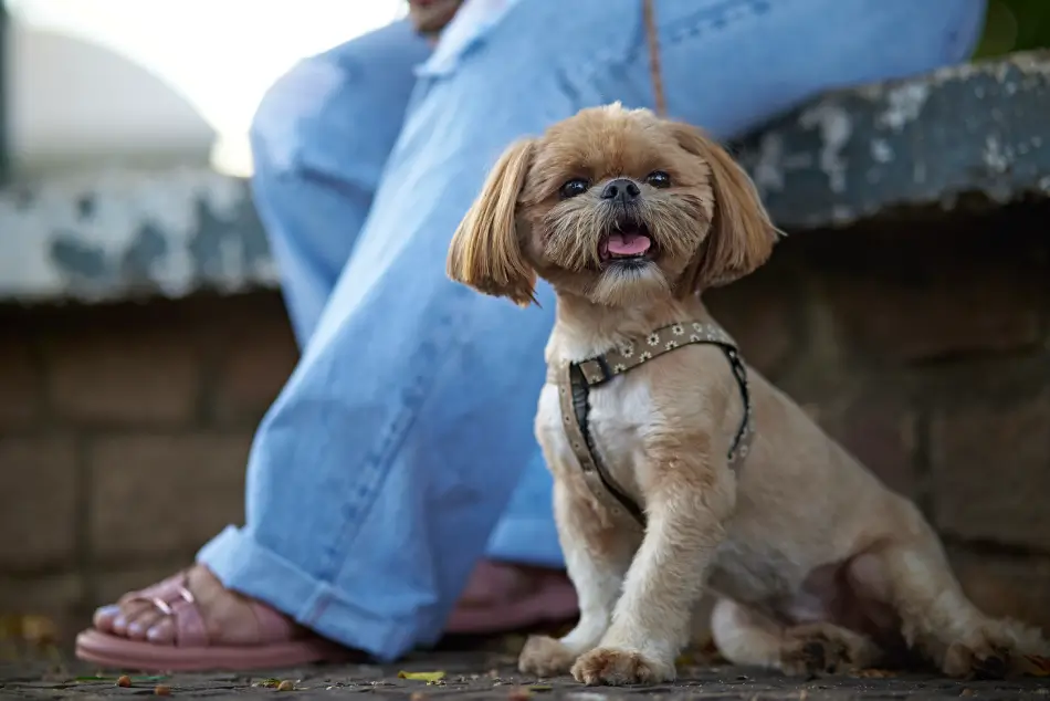
In a suburb near a large city, there lived a Shih Tzu named Barney. Barney was well-liked due to his friendly nature, but he struggled with a condition called macropalpebral fissure syndrome, which caused his eyelid openings to be abnormally large. This left his eyes prone to irritation because he could not fully blink, exposing his corneas to the harsh air.

Despite his condition, Barney and his owner, Mrs. Thistlewood, continued their daily walks in the neighborhood. Concerned about Barney’s discomfort, Mrs. Thistlewood consulted Dr. Hammond, a local veterinary ophthalmologist known for combining traditional practices with modern techniques. Dr. Hammond recommended a surgical procedure called medial canthoplasty to better protect Barney’s eyes by reshaping his eyelids.

The community valued Barney not just as a pet but as a local figure, and his surgery was a significant event. On the day of the operation, the neighborhood awaited news anxiously. The surgery went well, and although Barney’s recovery required time and he had to wear a “cone” or Elizabethan collar for a while, his tail soon showed his usual spirit.

Recovery progressed well, and before long, Barney returned to his regular activities with improved vision and more confidence in his steps.

Introduction

Large eyelid (see “eyelids”) openings in dogs can cause trouble. From Pugs to Great Danes, dog eyes are as varied as dog breeds are. Conformation of eyelids (palpebra) and eyelid openings (palpebral fissure) likewise can vary. 

Both shallow and deep orbits can be associated with abnormally large eyelid openings (macroblepharon, or macropalpebral fissure). Dogs with shallow orbits (see “orbit”) such as Pugs, Shih Tzus, and Pekingese are breeds that were created by humans to have the cute appeal of a wide-eyed infant. Unfortunately, the shorter the nose, the shallower the eye socket, and the more prominent the eye. Usually these breeds also have unusually large, round eyelid openings. The result is macropalpebral fissure syndrome. The hallmarks of this condition:

  • Dogs that cannot blink their eyelids effectively (“lagophthalmos”). Most of these dogs can do small incomplete blinks, in which the eyelids do not close and the center of the cornea is constantly exposed to air. Dogs that sleep with their eyelids partially open. The center of the cornea is constantly exposed to air. A desensitization of the constantly exposed corneas, so that the dog feels less of an urge to blink and does not squint or show other signs of pain as readily if the eye is injured.
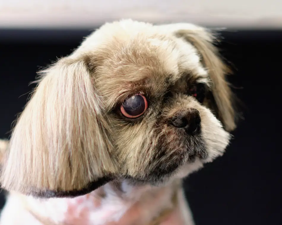
  • A prominent fold of skin over the nose (“nasal fold”). The hair on the nasal fold can rub on the surface of the eye (“nasal fold trichiasis”). Large nasal folds can also interfere with vision. Abnormal hair growing in the inside corners of the eyes, which is in constant contact with the corneal surface (“medial trichiasis”). This is especially common in Shih Tzus. The skin on the inside corner of the eyes rolls in (“medial entropion”), so that the hair on the surface of the skin also rubs on the cornea, causing irritation and increased rubbing of the face.
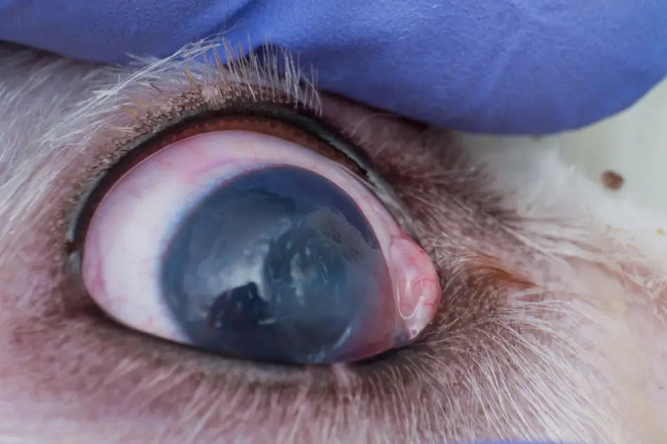
A high risk for either or both eyes to proptose (“pop” out of the eye socket) secondary to trauma. Proptosis often leads to blindness and sometimes the affected eye needs to be removed. A high risk for painful corneal ulcers to occur, and potential rupture and loss of the eye if the ulcer perforates. If the ulcer is severe, surgery can be performed to graft tissue into the corneal defect.

  • A high risk for eye trauma. Dogs with large eyelid openings and prominent eyes often love to rub their faces on the carpet and on furniture, damaging the eyes. This is particularly true in Shih Tzus, where excessive facial hair irritates the eyes if not kept clipped short. Additionally, if these dogs “play” with cats that don’t want to play, cat claw injuries commonly occur — the dog’s bulging, shiny eyes are easy targets for the cat.

Dogs with Macropalpebral Fissure Syndrome are often uncomfortable, and are at significant risk for blindness due to a film-like scar tissue forming on the corneas. Some dogs are blind before they are 5 years old.

Additional Complications
Many of these dogs also develop clinically dry eye, where their tear production and/or tearfilm (see “tear film”) quality is poor. If the eyes are dry in addition to the above mentioned problems, the dog is at extremely high risk of becoming blind from corneal pigmentation and scar tissue developing. Often, the corneal pigmentation is not noticed until it is quite advanced, as it is a dark brown color and blends in with the natural brown color of the iris.

Dogs will not begin to bump into objects until they have lost most of their vision– thus, if you notice a “film” on the surface of your pet’s eyes but your pet is visual, this does not necessarily mean that your dog’s vision is normal, and it is a good idea for your dog’s eyes to be examined by a veterinarian.

Additionally, English Bulldogs, Shih Tzus, and Pugs are prone to developing distichiasis, which are abnormal hairs that grow from the oil glands in the eyelids. These hairs, or “distichia”, can be located anywhere along the lid margin, and can be quite stiff and irritating. Sometimes they can actually grow on the inner surface of the eyelid, rubbing the eyeball like a sharp little needle. These inward-growing hairs are called ectopic cilia and they can be quite painful. See Canid Eyelid Diseases.

Advertisement

Treatment For Macropalpebral Fissure Syndrome
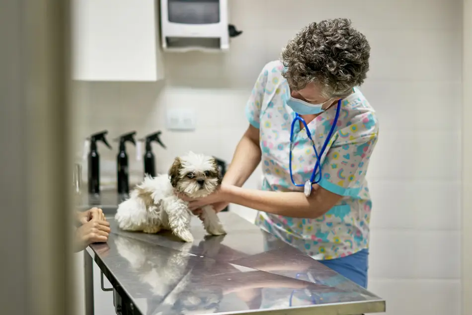
Dogs with macropalpebral fissure syndrome are best treated surgically, with a plastic surgery called medial canthoplasty, performed by a board-certified veterinary ophthalmologist. In this surgery, the inside corners of the eyelids are permanently sutured closed, effectively narrowing the eyelid openings and making them more almond-shaped. Additionally, the abnormal hairy tissue on the inside corners of the eyes is removed with this surgery, and the medial entropion is corrected.

This surgery does change the appearance of the dog’s face, as well it should. The patient needs to blink better and have less eye exposure to air, and to do this the eyelid openings must be smaller. An added benefit of this surgery is the reduced likelihood that the eye(s) could be proptosed secondary to trauma. Medial canthoplasty surgery does not always prevent future eye problems, but it can greatly reduce the frequency and severity of problems.

The first step in determining if your pet would benefit from medial canthoplasty surgery is to have a comprehensive ophthalmic examination by a veterinary ophthalmologist.  If your pet also has a dry eye disease, lifetime medical treatment may be required, in addition to medial canthoplasty surgery. If your pet also has either distichiasis or ectopic cilia, surgery to correct these problems will also be discussed and possibly recommended.

Macropalpebral fissure syndrome is a common condition that often requires both medical and surgical intervention, but with proper treatment, your dog can lead a long, happy life. Please ask for a referral to a veterinary ophthalmologist to schedule your pet for evaluation of this potentially blinding disease.
Terri L. McCalla

By Terri L. McCalla BSc, DVM, MS, Diplomate ACVO

Dr. Terri McCalla Dr. McCalla received a BS in Animal Health Science from the University of Arizona, followed by her DVM degrees from both Oregon and Washington State Universities in 1984.  She completed a 3 year residency in Comparative Ophthalmology from the University of Missouri and became board-certified in the American College of Veterinary Ophthalmologists (ACVO) in 1989.


🙀

This image may be disturbing to some viewers.

Fig 2. Close up of the eye of a dog with a deep corneal ulcer being held open by a blue gloved veterinarian.
Folds of skin that cover and protect the eye; they also spread tear film evenly over the eye surface when blinking.

Referring to the eyelids.

Also known as euryblepharon. Abnormally long eyelids which result in an abnormally large palpebral fissure (eyelid opening).

The bony socket that contains the eyeball and its associated muscles, nerves, and blood vessels. Damage to the orbital structure can affect eye movement, vision, and could potentially lead to loss of the eye.

A congenital condition characterized by overly prominent eyes and large eyelid openings, which can lead to increased corneal exposure and potential ocular complications. Common in brachycephalic breeds such as : (dogs) Pug, French Bulldog, Shih Tzu, and Pekingese; and (cats) Persian, Himalayan, and Exotic Shorthair.

Incomplete closure of the eyelids that can result in constant exposure and desensitization of the corneas. This can lead to corneal drying, ulceration, and scarring.

The transparent or clear tissue that covers the front part of the eye that includes the iris, pupil, and anterior chamber and bounded by the limbus. It functions to protect the inside of the eye from dirt, germs, and some ultraviolet light and helps to focus light.

A characteristic wrinkle of skin around the nose commonly present in short-nosed (brachycephalic) dog and cat breeds.

Occurs when hair covering a prominent nasal skin fold rubs directly on the eye, potentially causing irritation and vision issues. If left untreated, it can lead to corneal damage, including ulceration and/or keratitis. Usually affects both eyes.

The presence of abnormal hair growth in the inner corners of a dog's eyes that continuously contacts and potentially irritates the cornea. Over time, this irritation can lead to corneal ulcers and other eye health issues.

A condition where the skin at the inner corner of a dog's eye rolls inward, causing hair on the skin to rub against and irritate the cornea. This chronic irritation can lead to corneal damage and eye infections.

The displacement of an eye out of its bony socket in the skull ("popped out"), typically caused by trauma. This is a medical emergency that often causes blindness and often necessitates surgical removal of the eye, especially if not corrected as soon as possible.

Physical displacement of the globe from the bony socket, typically caused by trauma. This is a medical emergency that can lead to blindnesss.

Sores or erosions on the clear outer surface of the eye (cornea), typically caused by injury, irritation, or bacterial infection, and may result in pain, redness, and sometimes, impaired vision.
KCS, keratoconjunctivitis sicca. Poor tear production and/or quality leading to a dry ocular surface that can result in discomfort, conjunctivitis, mucoid discharge, keratitis with or without corneal ulcers, corneal and/or conjunctival pigmentation, and potential blindness.

A multi-layered film of fluids covering the eye surface that provides lubrication, prevents infection, and helps maintain clear vision. Disruption of this film can lead to dry eye syndrome, a common condition in many animals.

Abnormal growth of hairs from the oil glands (meibomian glands) of the eyelids, where these extra hairs may touch the surface of the eye, causing discomfort, eye irritation, and potentially leading to corneal damage or ulcers.

Extra eyelashes that grow to exit the eyelids at the meibomian gland openings. These hairs often cause irritation to the eye.

Tiny abnormal hairs that grow from the inner surface of the eyelid, pointing inward towards the eye. Ectopic cilia often cause significant pain and corneal ulcers, and can impair vision. Most common in young dogs.

A surgical procedure used to treat Macropalpebral Fissure Syndrome by narrowing the eyelid openings to reduce corneal exposure and correcting other eyelid changes causing irritation.

A veterinarian specialized in eye care who has passed rigorous exams to ensure a high level of expertise in the field of eye care and vision.

Advertisement

🙀

This image may be disturbing to some viewers.

Fig 2. Close up of the eye of a dog with a deep corneal ulcer being held open by a blue gloved veterinarian.
Folds of skin that cover and protect the eye; they also spread tear film evenly over the eye surface when blinking.

Referring to the eyelids.

Also known as euryblepharon. Abnormally long eyelids which result in an abnormally large palpebral fissure (eyelid opening).

The bony socket that contains the eyeball and its associated muscles, nerves, and blood vessels. Damage to the orbital structure can affect eye movement, vision, and could potentially lead to loss of the eye.

A congenital condition characterized by overly prominent eyes and large eyelid openings, which can lead to increased corneal exposure and potential ocular complications. Common in brachycephalic breeds such as : (dogs) Pug, French Bulldog, Shih Tzu, and Pekingese; and (cats) Persian, Himalayan, and Exotic Shorthair.

Incomplete closure of the eyelids that can result in constant exposure and desensitization of the corneas. This can lead to corneal drying, ulceration, and scarring.

The transparent or clear tissue that covers the front part of the eye that includes the iris, pupil, and anterior chamber and bounded by the limbus. It functions to protect the inside of the eye from dirt, germs, and some ultraviolet light and helps to focus light.

A characteristic wrinkle of skin around the nose commonly present in short-nosed (brachycephalic) dog and cat breeds.

Occurs when hair covering a prominent nasal skin fold rubs directly on the eye, potentially causing irritation and vision issues. If left untreated, it can lead to corneal damage, including ulceration and/or keratitis. Usually affects both eyes.

The presence of abnormal hair growth in the inner corners of a dog's eyes that continuously contacts and potentially irritates the cornea. Over time, this irritation can lead to corneal ulcers and other eye health issues.

A condition where the skin at the inner corner of a dog's eye rolls inward, causing hair on the skin to rub against and irritate the cornea. This chronic irritation can lead to corneal damage and eye infections.

The displacement of an eye out of its bony socket in the skull ("popped out"), typically caused by trauma. This is a medical emergency that often causes blindness and often necessitates surgical removal of the eye, especially if not corrected as soon as possible.

Physical displacement of the globe from the bony socket, typically caused by trauma. This is a medical emergency that can lead to blindnesss.

Sores or erosions on the clear outer surface of the eye (cornea), typically caused by injury, irritation, or bacterial infection, and may result in pain, redness, and sometimes, impaired vision.
KCS, keratoconjunctivitis sicca. Poor tear production and/or quality leading to a dry ocular surface that can result in discomfort, conjunctivitis, mucoid discharge, keratitis with or without corneal ulcers, corneal and/or conjunctival pigmentation, and potential blindness.

A multi-layered film of fluids covering the eye surface that provides lubrication, prevents infection, and helps maintain clear vision. Disruption of this film can lead to dry eye syndrome, a common condition in many animals.

Abnormal growth of hairs from the oil glands (meibomian glands) of the eyelids, where these extra hairs may touch the surface of the eye, causing discomfort, eye irritation, and potentially leading to corneal damage or ulcers.

Extra eyelashes that grow to exit the eyelids at the meibomian gland openings. These hairs often cause irritation to the eye.

Tiny abnormal hairs that grow from the inner surface of the eyelid, pointing inward towards the eye. Ectopic cilia often cause significant pain and corneal ulcers, and can impair vision. Most common in young dogs.

A surgical procedure used to treat Macropalpebral Fissure Syndrome by narrowing the eyelid openings to reduce corneal exposure and correcting other eyelid changes causing irritation.

A veterinarian specialized in eye care who has passed rigorous exams to ensure a high level of expertise in the field of eye care and vision.

Already have an account?

Two ways to read this article:

By continuing, you agree to the Privacy Policy, Terms of Use